Science meets art: Maree Clarke and the Melbourne Histology Platform
The study of microscopic anatomy – histology – is an inherently visual process, using tissue sectioning and staining procedures to reveal the presence of molecules and the morphology of cells and tissues of interest to pathologists and biological researchers. But to multidisciplinary visual artist Maree Clarke, the potential of collaborating with the Melbourne Histology Platform reaches far beyond scientific discovery.
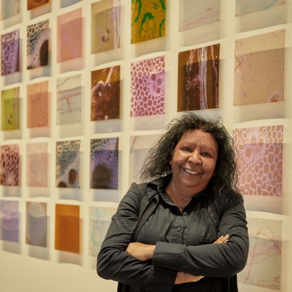
Maree Clarke's latest work on display at ACCA, now you see me: seeing the invisible #1, was produced by applying the science of histology to the water reeds which have featured in her work for over a decade. Photo: Casey Horsfield/ACCA
Curated by Jessica Clark, the exhibition Between Waves at the Australian Centre for Contemporary Art presents the work of ten artists and collectives whose work aims to illuminate interconnected shapeshifting ecologies within, beyond and between what can be seen.
Maree Clarke has been working with water reeds as part of her artistic practice for over a decade, including making a 50m river reed necklace. But for her contribution to Between Waves, she wanted to move from supersized to the opposite end of the size scale. “The site where ACCA stands was once expansive wetlands that would have been filled with river reeds […] in response to place, I started wondering about the micro systems, in what is now a built environment. The ecosystems of wetland areas, seen and unseen”.
Maree’s search for guidance into the microscopic world led her to the Melbourne Histology Platform, the University of Melbourne’s research platform (based in the School of Biomedical Sciences) that assists researchers to prepare biological tissue samples to investigate their microscopic anatomy using optical microscopy.
“The challenge for the team wasn’t so much about an unusual sample,” says evolutionary biologist and MHP's Platform Manager Dr Chris Freelance. “Usually, a researcher brings in samples wanting to visualise a particular tissue or cellular structure to answer a research question, and the samples then undergo a staining process will highlight that particular biological target with a particular colour based on chemical reactions. But Maree’s primary interest was about seeing cellular structures in a visually compelling way rather than looking for something specific. This meant starting with a range of stains typically used for plant histology, seeing what Maree got out of it and then how we could tweak things from there”.
After an initial meeting including Dr Paul McMillan from the Biological Optical Microscopy Platform, MHP’s professional histologists, Laura Leone and Lisa Foster, took the river reed stems and roots through the steps of processing them to preserve morphology, embedding them in paraffin wax, cutting them into 2 micron-thick cross-sections placed onto microscope slides, and finally staining them with three different protocols. To everyone’s delight, the initial results were more than Maree had imagined. “I love it! It's one of my new favourite things”.
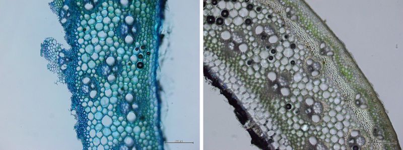 Stained with toluidine blue (left) and left unstained (right), seeing river reeds at the microscopic scale gives a very different view to what you see with the naked eye. Photos: Maree Clarke
Following some tweaks and histological preparation of some additional river reed sections, she had all the slides she wanted. After several hours with Freelance learning how to use the microscope to perform brightfield, darkfield, and polarised light microscopy, she captured a series of hundreds of images which she later turned into her piece now you see me: seeing the invisible #1 – a collection of 297 micrographs.
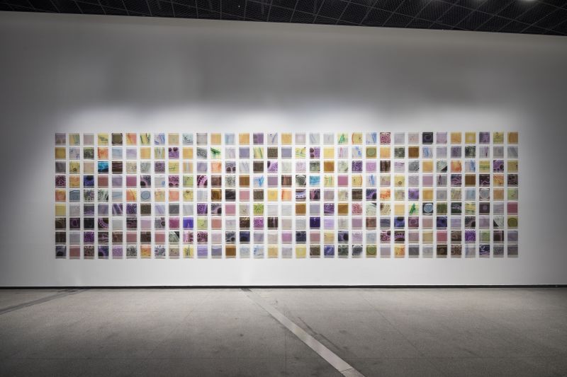
Maree’s piece for Between Waves, now you see me: seeing the invisible #1, is a collection of 297 micrographs printed on acetate. Photo: Andrew Curtis.
“This was an amazing project for MHP to be involved with”, says Freelance. “Histology and light microscopy are very much visual processes as we’re discerning the composition of biological samples based on the colours that result from chemical staining reactions. This has been a fantastic collaboration helping someone like Maree integrate histology into her art practice to bring the microscopic world to a broader audience. It’s a great example of science and art meshing seamlessly”.
A biologist might look at the stained river reed sections and, based on morphology and the staining reactions, see vascular bundles containing xylem vessel cells with thick cell walls composed of lignin, or areas of parenchyma tissue with thin cellulose-based cell walls. But to Maree, that view down the microscope depicts much more. “I saw unseen worlds that look like so many different things; nebulas, weavings, eyes, seascapes, brushstrokes, cobwebs, bubbles, chicken wire, coastlines, whole galaxies and more, and that’s a pretty mind-blowing experience”.
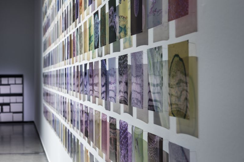
now you see me: seeing the invisible #1 is a collection of 297 micrographs printed on acetate. Photo: Andrew Curtis.
I have loved being able to go into the Histology department at Melbourne Uni, to meet these incredible people to make this new body of work was just amazing! And for people to see just how beautiful the microscopic river reed that was revealed to me through that lens.
The exhibition Between Waves is on show at ACCA until 3rd September 2023.
An extension of the exhibition, Maree Clarke’s piece now you see me: seeing the invisible #2 will be displayed on the screens of Melbourne’s Fed Square from 21st August – 3rd September 2023.
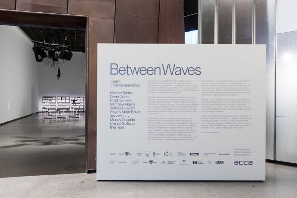
The exhibition Between Waves is on show until 3rd September 2023. Photo: Andrew Curtis.