The role of glial cells in retinal vascular disease
Project Details
The retina contains two major classes of glia, Müller cells and astrocytes, and both are integral to the way the retina functions. Both types of glial cells play an important role in providing metabolic substrates to neurons, deactivation and recycling of neurotransmitters and maintain the ion balance of the retina. Müller cells and astrocytes also maintain the integrity of the blood-retinal barrier and regulate blood vessels in response to neuronal activity. Dysfunction in retinal glia has been implicated in many retinal diseases, including Diabetic retinopathy and Retinopathy of Prematurity. We and others have reported numerous changes in Müller cell and astrocyte function at various times during the progression of diabetes, including changes in vascular endothelial growth factor (VEGF) a major stimulant for angiogenesis of retinal blood vessels, and alterations in astrocyte number and communication. This project will use immunohistochemical, molecular and cell-based technologies to examine the changes in Müller cells and astrocytes that lead to altered vascular function in pre-clinical models of diabetic retinopathy and retinopathy of prematurity.
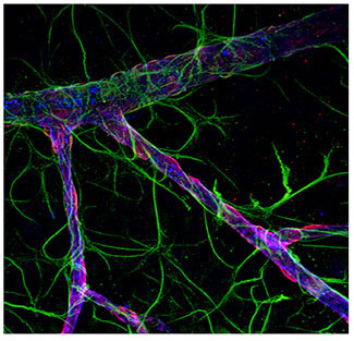
Figure 1: Astrocytes are intimately linked to the function of the inner retinal vasculature.
A flat-mounted retina labelled for astrocytes (green) and retinal vasculature (pink).
Researchers
Dr Andrew Jobling, Senior Research Officer
Funding
Australian Research Council