Why giraffes have spots
A/Prof Quentin Fogg & Prof Ian Taylor research the link between the purpose of giraffe patches and applications in plastic surgery.
This article was first published on Pursuit. Read the original article.
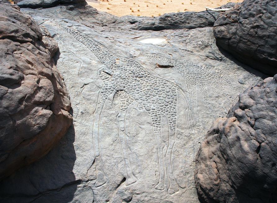
Giraffe spots have interested researchers and natural historians for hundreds of years, and indeed humankind for much longer.
In fact, they are a key feature in early rock carvings, including life-size engravings found in the Aïr Mountains of the Sahara dating back 6,000 to 8,000 years.
Now, thanks to a reanalysis of studies by the internationally renowned plastic and reconstructive surgeon Professor Ian Taylor (Dept Anatomy & Physiology) and his team in the 1990s – which involved dissection and injecting the patches with specially produced contrast media – we can provide the first anatomical proof of why a giraffe’s spots exist.
Published in Plastic Reconstructive Surgery, the new paper involving A/Prof Quentin Fogg and Professor Taylor explains the remarkable link between the purpose of the patches – almost certainly unique to the giraffe – and applications in plastic surgery.
A NEW UNDERSTANDING
Rather than being uniformly dark, it appears that evolution has rearranged the giraffe’s skin pigmentation to combine camouflage with thermoregulation.
For the past 15 years, a basic model of the blood supply has been used to describe how giraffe patches relate to temperature control.
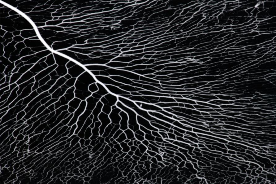
"Our new analysis of Professor Taylor’s anatomical work reveals that, in fact, each patch has its own, single, unique, central artery," say Quentin Fogg.
The artery then sends long radiating branches in a broad, delicate and beautiful fan to fill the entire patch.
Remarkably, on reaching the pale skin boundary these branches are connected by shunts to large veins that encircle the patches and connect to adjacent patch arteries. This architecture allows large volumes of heated blood to be dumped into these large veins or into adjacent cooler patches.
Known as an angiosome, this blood vessel architecture occurs where a certain section of the body – that can include a 3D combination of skin, fat, bone and muscle – is supplied with blood by a key source artery and drained by other specific veins.
Professor Taylor and his team mapped the entire human body into over 300 angiosome territories over his 50 years of anatomical research, a body of work that has given rise to the field of microvascular free tissue transfer (free flaps) used in every reconstructive plastic surgery unit in the world.
This study has revealed that the giraffe’s angiosomes spots are currently the most detailed and specialised system known for the rapid shunting of high volumes of warm blood discovered to date.
TEMPERATURE REGULATION
As described by Professor Taylor, like a traffic light, this unique vascular anatomy of the giraffe patches allows them to ‘switch on or off’ to expel or retain heat shunted directly to the lungs from where the heat is most efficiently expelled.
This ingenious mechanism saves muscles, organs and other structures from dealing with the high temperatures during the day and can strategically redeploy blood with the greatest efficiency – an evolutionary benefit that can mean the difference between life and death in the natural habitat of the giraffe.
At night, when temperatures plummet, the shunting vessels close to keep the warmer blood around the muscles and organs, helping the giraffe maintain its temperature.

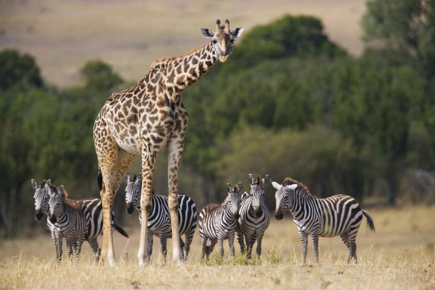
In this study, we also reanalysed the zebra, African hunting dog and jaguar – all have similar markings – but none have an angiosome pattern similar to the giraffe that matches their pigmented areas.
So, these spots in the giraffe have almost certainly evolved as a truly unique feature.
ENVIRONMENT AND EVOLUTION
The exciting identification of shunts in the giraffe has raised many other questions that should be explored.
For example, does the okapi – the closest evolutionary relative of the giraffe – also have skin blood vessel architecture that matches its dark skin markings?
Or has this angiosome pattern not developed in the okapi because of its reclusive nature, where it is seldom seen emerging from dense shaded foliage?
Are the blue rings of an octopus also discreet angiosome patches with shunts that can switch on and off to deter its predators?
And what is it about the nerve and blood systems of the chameleon skin that permit colour change to match not only its thermal but visual environment? A close consideration of these extreme anatomical adaptions will help inform our understanding of human anatomy and physiology.
IMPLICATIONS FOR PLASTIC SURGERY
This revelation of a shunting mechanism in the giraffe to divert blood rapidly from arteries to veins may be important in understanding human physiology and pathology.
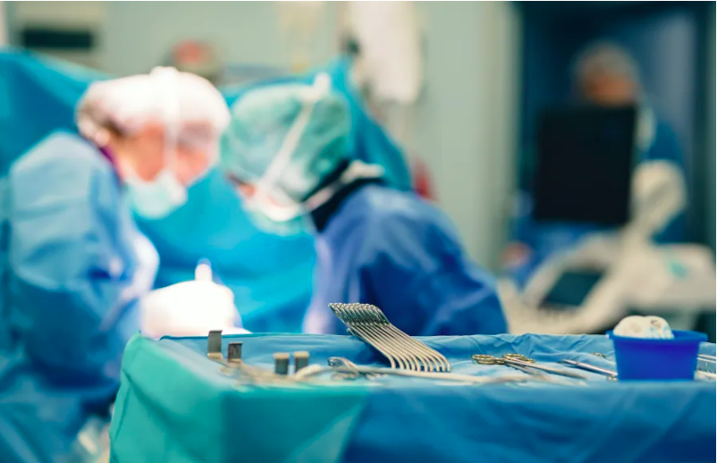
For example, the angiosome blood supply pattern is used daily in plastic surgery.
Surgeons use knowledge of which artery supplies what area of tissue in skin and underlying tissue transplants. In cases like breast reconstruction or large wound repair, the blood supply to the transplant is reconnected using microsurgery.
In these operations, do shunts play an important role in adjusting flow where there may be a sudden influx of high-pressure blood from a large to a smaller artery when the clamps are released?
The giraffe’s model of blood use to control temperature could also have unique applications in plastic surgery, especially when considering the exposed and vulnerable skin of the face.
Professor Taylor has also recently demonstrated unique shunt connectivity between blood vessels around the eye.
This paper demonstrates how important these systems can be for temperature control, especially for the most habitually exposed parts of our bodies and how cosmetic fillers can accidentally reach the eye, causing blindness.
"Our aim is for these results to inform ongoing investigations into vascular control in humans, in turn creating more decisive and reproducible results for our patients," Quentin says.
FUTURE RESEARCH
The next major step is a search for arteriovenous (AV) shunts in key parts of the human body where they may be important for maintaining body homeostasis – including sudden changes in blood pressure or diversion of blood toxins.
Of special interest are the limbs, where AV shunts – larger than the tiny ones documented in the hairless skin of the hands and feet – must exist but are not clearly documented.
Once identified, they could inform the modification and development of plastic surgery techniques.
The University’s Melbourne Academy of Surgical Anatomy (MASA) plans to bring together these new ideas and perspectives to influence the practice of surgery in the future.
"We are grateful to the giraffe’s spots for helping us understand more about our own anatomy, while we study the origin of its amazing markings," Quentin concluded.
Researchers from The Department of Plastic Surgery at the Royal Melbourne Hospital; Department of Anatomy and Physiology, the University of Melbourne; Craniofacial and Plastic Surgery Research Unit at The Prince of Wales and Sydney Children’s Hospital, University of New South Wales; St Vincent’s Institute for Medical Research and Division of Plastic Surgery, Dalhousie University, Halifax, NS, Canada also contributed to this paper.