Going Viral: Exploring Viruses in Virtual Reality
Master of Biomedical Science students see real-world applications of virtual reality (VR) at cutting-edge workshop held in the Digital Learning Hub.
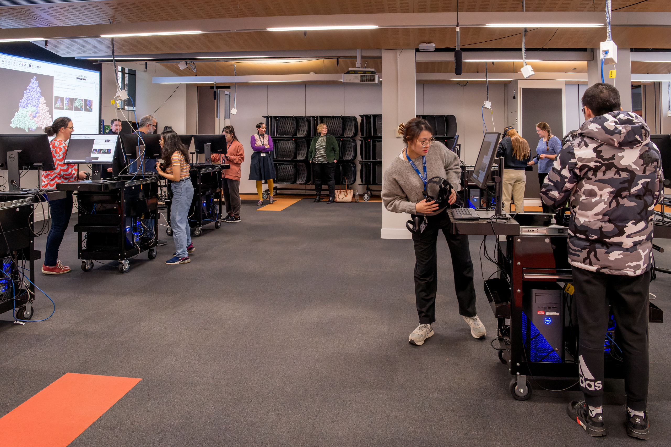
Students get ready to don headsets and explore viruses in VR
In an innovative learning experience, Master of Biomedical Science students in the Defence and Disease: Frontier Technologies subject experienced life as a modern virologist. The exciting new workshop created by Dr Jason Roberts (Royal Melbourne Hospital & Peter Doherty Institute for Infection and Immunity) and Dr Sarah Londrigan, Senior Lecturer Virology and coordinator of the Defence and Disease subject (Department of Microbiology & Immunology), let students visualise real-world applications of VR used in the intersecting disciplines of public health and basic research.
The workshop, led by Dr Jason Roberts and Ms Jamie Mumford (Electron Microscopy and Structural Virology Laboratory at the Doherty Institute), let students embark on a journey of scientific exploration that merged technology, public health and academia like never before.
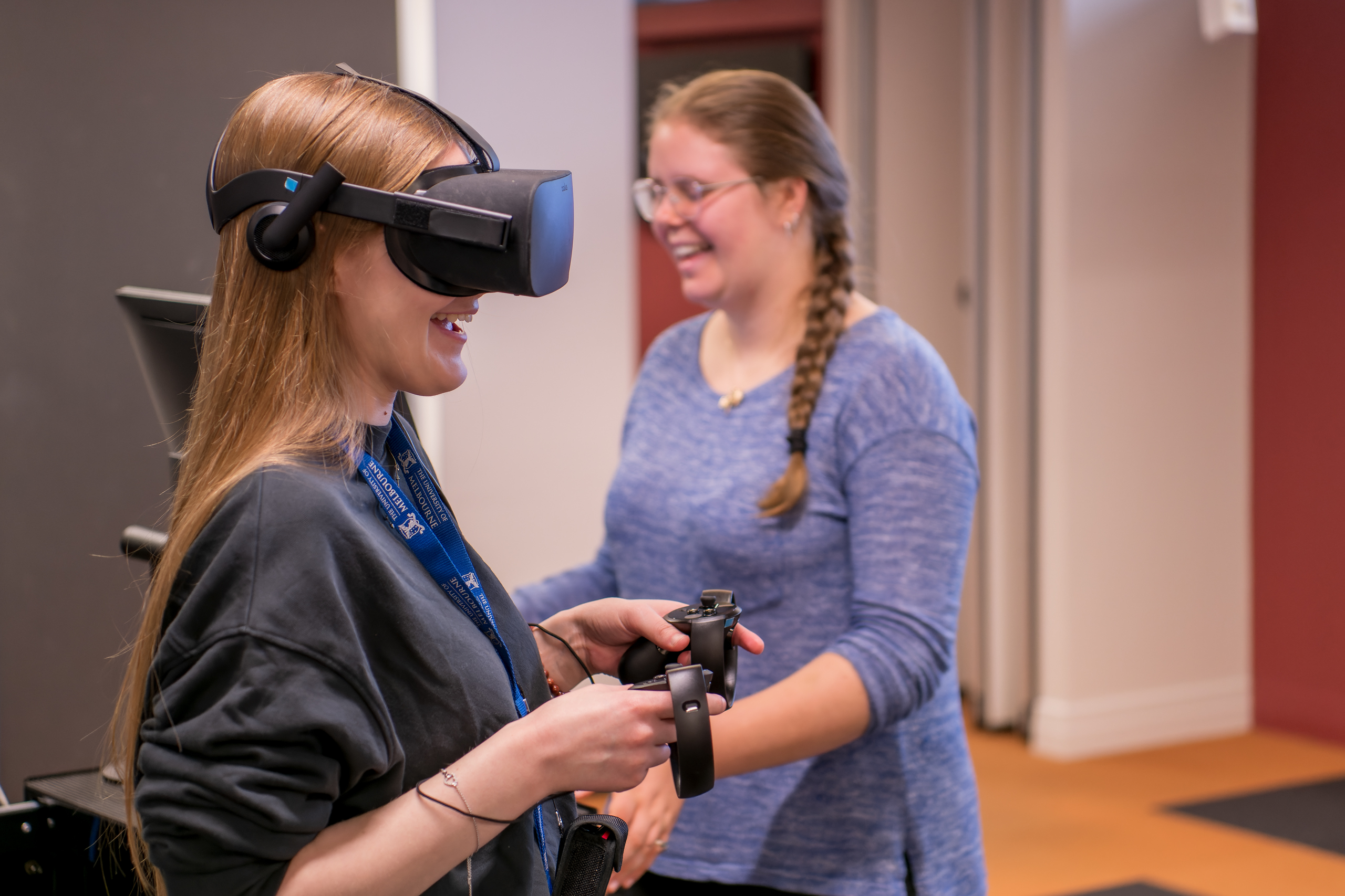
They immersed themselves (safely!) inside an infectious virus particle through virtual reality (VR) in the Digital Learning Hub - and found out how a modern virologist works in virtual reality (VR), using samples from Dr Roberts own published work.
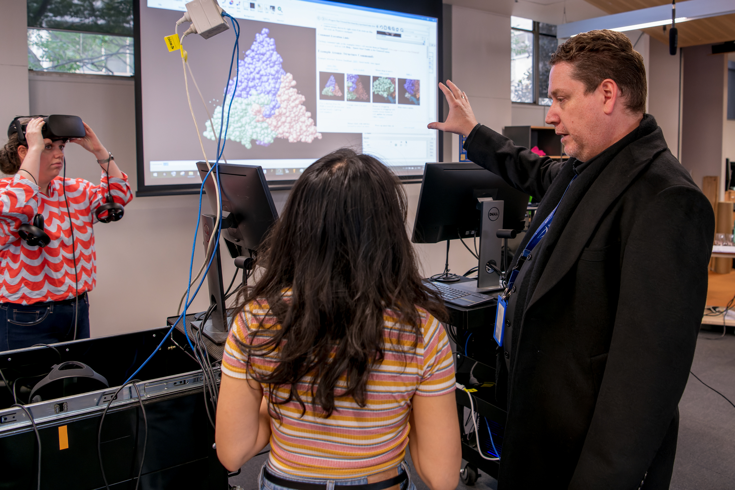
Dr Jason Roberts (right) teaches students how to manipulate virus structures in virtual reality
Under Dr. Roberts' expert guidance, the students delved into the intricate world of viruses by examining the three-dimensional structure of the insect virus black beetle nodavirus in VR using ChimeraX. They learned to navigate through three-dimensional structures and how to manipulate various display settings to gain a comprehensive understanding of the viral anatomy. Additionally, the students practiced aligning cryo-electron micrographs with atomic models, a crucial aspect of advanced virological research.
The ability to rapidly derive and visualise electron microscopy data in 3D virtual reality space, particularly during events of significant public health impact, adds a whole new dimension to the identification and morphological characterisation of pathogens and related atomic structures. Combined with advances in artificial intelligence, the future of imaging in relation to pathogen identification, reference and research is immensely exciting.
Once the students had familiarized themselves with the intricacies of working with viruses in a virtual environment, they turned their attention to a sample of Mpox-infected cellular material provided by Dr. Roberts. Unlike traditional 2-dimensional screens, the VR technology allowed the students to freely explore the entire slice of infected cells, enabling them to observe the distinctive dumbbell-like structures within the Mpox virions, that serve as a diagnostic hallmark of poxvirus infection.
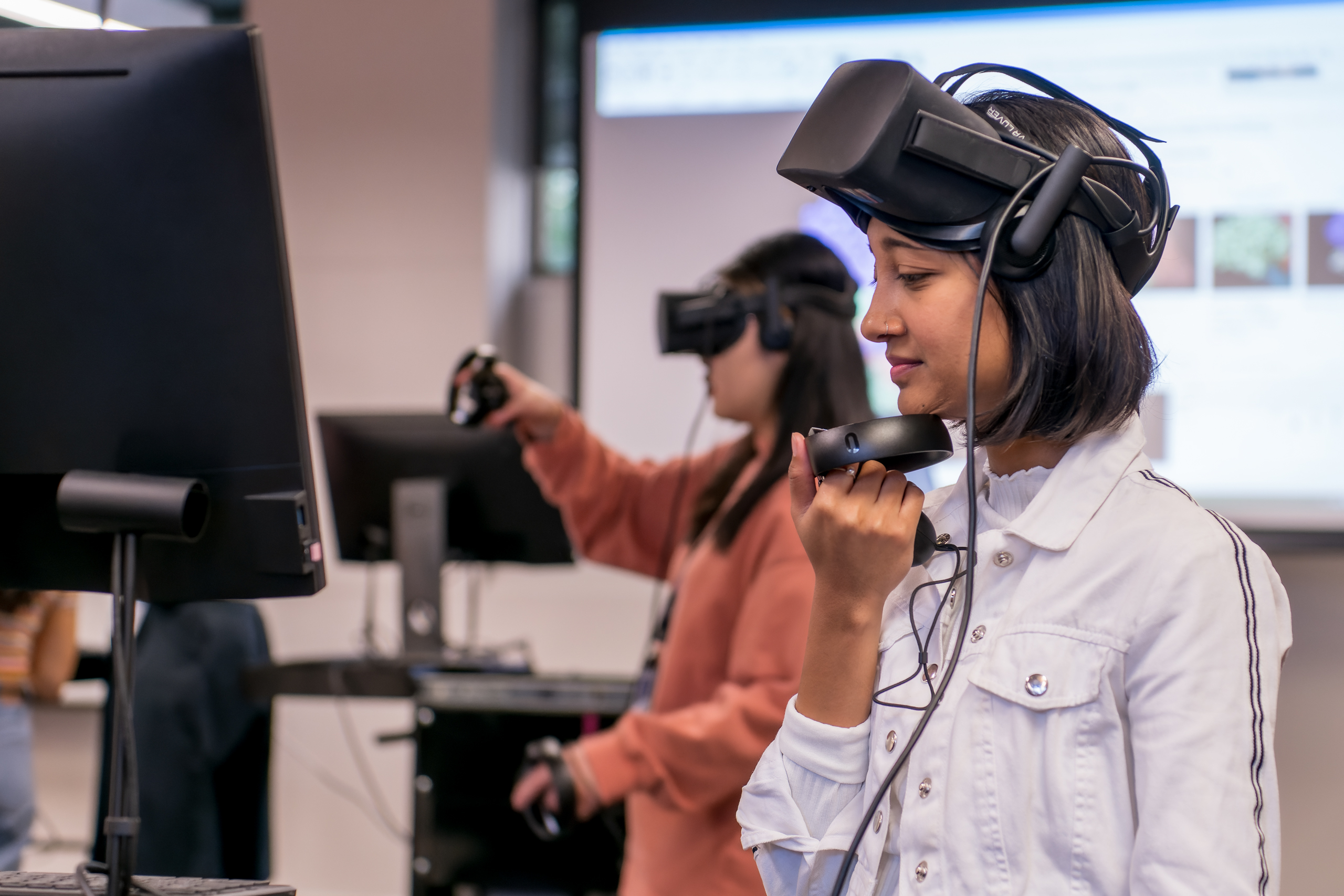
A student considers the next step in the workshop.
The use of VR in the subject Defence & Disease: Frontier Technologies (BMSC90019), coordinated by Dr Sarah Londrigan, Dr Jessica Welch and A/Prof Odilia Wijburg in the School of Biomedical Sciences, not only provided a realistic and engaging experience, but also offered the students a unique perspective on the subject matter. By transcending the limitations of conventional learning methods, this immersive VR adventure granted the students a sense of freedom to examine infectious diseases in a more comprehensive and interactive manner, using the same technologies that experts in the field use daily.
This subject introduces students to a range of specialised and emerging research techniques and technologies and applies them to understanding contemporary research problems that intersect with public health responses.
— Dr Sarah Londrigan
This article was originally published by the Digital Learning Hub on 2 June 2023.