Pathology undergraduates visit MHP for third year running
For the third consecutive year, the Melbourne Histology Platform has proudly hosted undergraduate pathology students for a hands-on look into the daily workflow of a histology laboratory.
The sessions for students from the undergraduate coursework subject PATH30002 Techniques for Investigation of Disease began with an overview of the core processes, from tissue fixation to paraffin processing and embedding. Students had the opportunity to test their dexterity using forceps and molten wax to embed human placental tissue, gaining a real feel for this critical technique.
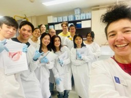
PATH30002 students (group #3) with MHP's Laboratory Coordinator Laura Leone.
They then observed paraffin sectioning using a microtome and had the chance to collect delicate 5-micron lung tissue sections onto glass slides. The next stop was automated Haematoxylin and Eosin (H&E) staining, where students were amazed by the precision of the stainer, particularly the delicate placement of coverslips, a task they’re used to doing manually in class!
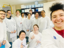
PATH30002 students (group #6) with Laura Leone.
At the microscope station, they explored the significance of general tissue morphology with the H&E stain and discussed how these observations are the first step in guiding further diagnostic investigations with special stains.
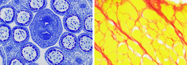
Toluidine blue stain of a flower bud (left) and picrosirius red stain of muscle tissue (right).
But the learning didn’t stop there. Students were introduced to the unique intersection of science and art where the patterns seen under the microscope inspire artistic expression beyond working hours. Histology, they discovered, is not only a science but also a source of visual wonder.
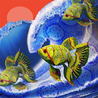 Histology-inspired digital artwork by Laura Leone.
-Article by Laura Leone