PATH30002 student visits to MHP
It’s always rewarding to see students engaged in discovering how a histology laboratory really operates. Recently, undergraduate pathology students had the opportunity to observe MHP's day-to-day workflow.
Microns and Mysteries: A Student’s Look Inside Histology
In April, undergraduate pathology students from the University's PATH30002 Techniques for Investigation of Disease subject had the opportunity to step inside the Melbourne Histology Platform and observe the day-to-day workflow in action.
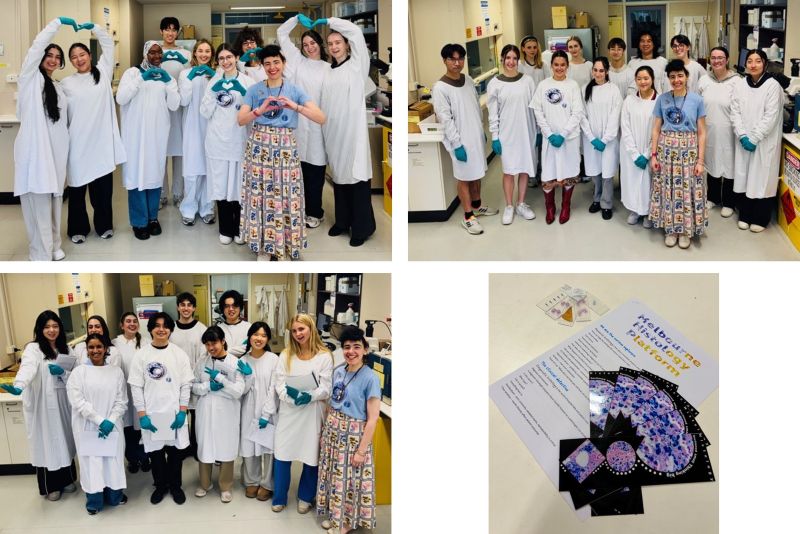
A key focus of the visit was understanding how research histology differs from diagnostic histology. In clinical settings, histology plays the role of a detective, working to deliver clear answers that guide patient diagnosis and treatment. In contrast, research histology is driven by curiosity. It’s less about immediate answers and more about asking new questions: What don’t we know yet, and how can we find out?
Despite these different aims, both areas share a common foundation. Core techniques such as tissue fixation, processing, embedding, and sectioning underpin all histological work. Students explored staining approaches from routine Haematoxylin and Eosin (H&E) through to special stains and immunohistochemistry alongside the microscopy skills and attention to detail essential for accurate interpretation.
Across three enthusiastic groups, students were introduced to the investigation of disease, beginning with an overview of core processes from tissue fixation through to paraffin embedding. A highlight for many was seeing ultra-thin paraffin sections cut on a microtome and having the chance to collect sections onto slides themselves.
We also explored how staining evolves alongside scientific discovery. Students learned about the importance of H&E as a foundation stain for assessing tissue morphology, and how it often leads to the use of more targeted techniques. For example, Perls’ Prussian blue stain is used to detect iron in tissue. Through research innovation, this method was further developed into a DAB-enhanced version, allowing scientists to detect subtle iron accumulation in the brain in conditions such as Alzheimer’s disease that would otherwise go unnoticed.
To round out the experience, students were shown the remarkable diversity of samples processed within our laboratory. From oysters and lizards to parasites and plant material such as leaves and bark, the range reflects the breadth and curiosity-driven nature of research histology.
It was a pleasure to host such inquisitive groups, and we look forward to inspiring the next generation of scientists to keep asking questions and exploring the unknown.
-
Text by Laura Leone.
University of Melbourne subject coordinators interested in MHP visits/sessions for their subjects in future years should contact the Platform Manager, Dr Chris Freelance.