Image competition to showcase Anatomy & Physiology research
Over 20 stunning entries received from academic, research staff and HDR students. View the winners here.
The images showcase exceptional creativity and the vibrant and diverse research underway across the Department's research themes: Metabolic and Cardiovascular Sciences; Muscle Biology; Sensory Neuroscience; Stem Cell and Developmental Biology and Systems Neuroscience.
The internal competition coordinated by the Department of Anatomy & Physiology's Research Committee was designed to source original scientific images to be used to visually enhance and elevate the Department's externally facing research profile.
Please join us in congratulating our winning entrants:
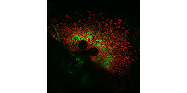
Stacey Keenan
- Theme: Metabolic and Cardiovascular Sciences
- Title: Lipid droplets and mitochondria in primary hepatocytes
- About: Image depicting an isolated primary hepatocyte, examining the mitochondria–lipid droplet interaction, stained with Mitotracker (red) and Bodipy (green) to visualize mitochondria and lipid droplets, respectively.
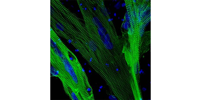
Dr Kevin Watt
- Theme: Muscle Biology
- Title: Human muscles
- About: The image show 3D human muscle tissues cultured for 7+ days. The muscle tissues are labelled for the structural protein Titin (green) showing the complex architecture of the muscle fibre and nuclei (blue). The generation of 3D human muscle tissues provides new capabilities to model complex acquired and inherited muscle diseases.
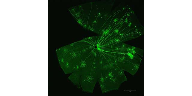
Dr Ursula Greferath
- Theme: Sensory Neuroscience
- Title: Thy1+ganglion cells
- About: Thy1-driven GFP fluorescence in ganglion cells of the mouse retina
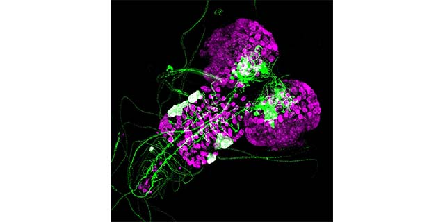
Edel Alvarez
- Theme: Stem Cell and Developmental Biology
- Title: Portrayals of Prospero
- About: Drosophila melanogaster larval brain with tumours. The knock-down of the neuronal-fate determinant prospero in Neural Stem Cells (NSC, in magenta) causes cells to fail to differentiate into neurons, leading to increased proliferation of NSC and ultimately tumour growth (in white). Additionally, we can see neuronal projections and axons in green.
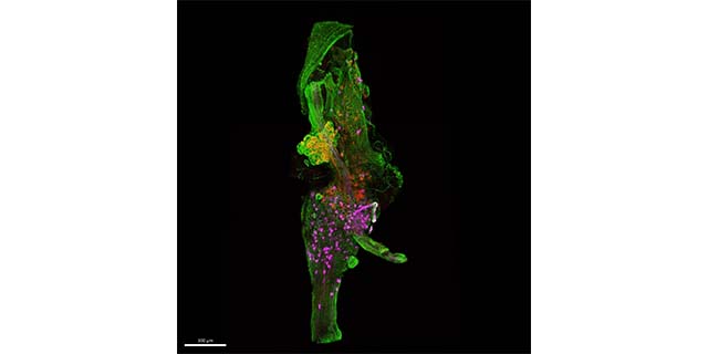
Dr Aung Kywe Moe
- Theme: Systems Neuroscience
- Title: A blooming bud
- About: This is a three-dimensional image of mouse vagal ganglia which have been labelled with three different markers of neurons, chemically treated to become transparent and imaged at a confocal microscope. The neurons and nerve fibres which are embryologically derived from a special group of cells called neural crest are labelled with green fluorescent protein. Two other subtypes of vagal neurons and their fibres are labelled with red and far red (pseudo-coloured as magenta here) fluorescent proteins.
With thank to A/Prof Kelly Smith, Dr Song Yao, Dr Helen Jiao who led the initiative and judged the competition.