Phenomics Australia Histopathology and Digital Slide Service
Welcome to the Phenomics Australia Histopathology and Slide Scanning Service website
Phenomics Australia Histopathology and Slide Scanning Service
Supported by Phenomics Australia
The Phenomics Australia Histopathology & Slide Scanning Service provides necropsy and histopathology services to all biomedical researchers across Australia for evaluating or phenotyping modified, treated, experimental or genetically engineered mice at all developmental stages.
A team of experienced medical, veterinary pathologists and mouse pathobiologists determine whether morphological changes or pathology identified are strain-related, age-related, incidental or a true diversion from normal. The mouse strains are therefore validated and utilised as models of human disease.
The Histopathology and Digital Slide Service is based at the Department of Anatomy and Physiology at the University of Melbourne and is supported by the Federal Government through the National Collaborative Research Infrastructure Strategy (NCRIS) initiative and by the University of Melbourne.
Our standardised procedures are designed to provide a thorough, first-line approach to assessing the entire animal or organs of interest.
Evaluations
- Professional commentary from medical and veterinary pathologists and mouse pathobiologists
- Necropsy and full-body histopathology (rodent)
- Semi-quantitative analysis-Tissue Scoring
- Mouse neonates and embryos
- Blood and bone marrow smears
- Terminal blood analysis
Request service here (iLab)
Imaging
- High-quality, high-speed brightfield slide scanning (slide digitization)
- Macroscopic/microscopic image documents accompanying all Histopathology reports
- Secure online access to all digitized images and histopathology reports
The Histopathology and Digital Slide Service provides a secure interface where users can login and interrogate their case files, reports and images.
To view your images access slides.
Tissue Scoring and Grading
Through experience and careful collaboration with our pathologists and researchers, our service has developed standardised protocols to enable a semi-quantitative approach to evaluate the effects of treatments. These protocols are unique to each tissue and can be project specific.
Data Curation
Secure online access to large format, interrogable histological images created utilising state-of-the-art image acquisition systems.
Access SlideCenter here
The Platform is a fee-for-service operation, open to all not-for-profit and corporate requests.Staff are always available to assist.
You may wish to explore the Phenomics Australia Voucher Scheme
Phenomics Australia offers a voucher scheme to facilitate and encourage access to one or more Phenomics Australia facilities. This scheme is intended to further reduce the cost of access to a specific capability
Complementary platform technologies/services:
Terms & Conditions
Read the terms and conditions (DOCX 32.1 KB)


![]()
Tina Cardamone
Service Manager
Email: t.cardamone@unimelb.edu.au
Professor Gary Hime
Phenomics Australia Histopathology and Digital Slide Service Unit Leader
Email: g.hime@unimelb.edu.au
General Email: histopathology-scanning@lists.unimelb.edu.au
Location:
Room E236,
Department of Anatomy and Physiology,
Medical School Building (Corner of Grattan Street and Royal Parade),
University of Melbourne
Follow us on LinkedIn


Emily Gracie

Veronika Purchase

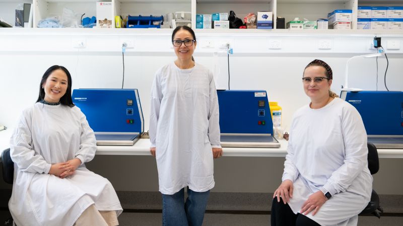
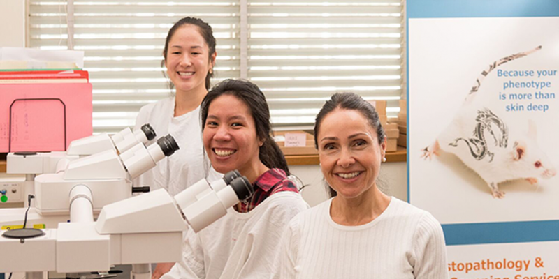
The Team
The Histopathology Team has the expertise to provide skilled evaluation of variant strains and has comprehensive knowledge of background strains, experience in mouse pathology, familiarity with mouse anatomy and understanding of unique mouse micromorphological features.
Consultant Pathologists
Phenomics Australia has access to leading diagnostic and academic Pathologist
Professor Catriona McLean
Medical Pathologist, Neuropathologist & Head of Department of Anatomical Pathology, Alfred Health, Melbourne, Victoria

Dr John Finnie
Senior Comparative Veterinary Pathologist, The University of Adelaide, South Australia

Phenomics Australia Histopathology and Digital Slide Scanning Service
Production of high-quality, large format digital slide images for further analysis and publication
Secure access to all your digital images through a user friendly interface- SlideCenter
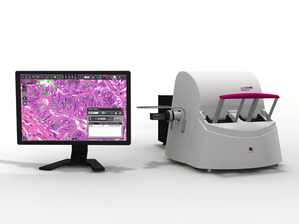
3D Histech Pannoramic ScanII Scanner
This is a high resolution slide scanning device capable of imaging standard stained microscope slides (brightfield). Digital images can be interrogated by the client with the free SlideViewer software https://www.3dhistech.com/software-downloads
(Note: software is only compatible with windows operating systems). Users have the option to either collect and copy their images or for a small fee access and view their images via the SlideCenter web interface
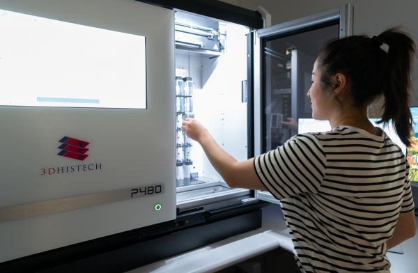
P480 Slide Scanner
The Pannoramic 480 DX RUO is an ultra high throughput volume scanner.
World class brightfield whole slide scanning
Fitted with 20x and 40x objectives
Up-to 480-slide capacity robotized loader
This instrument is part of Phenomics Australia and is available to University of Melbourne and external users.
Digital images can be interrogated using the free 3D Histech SlideViewer software or through the online slide manager SlideCenter.
The P480 Slide Scanner is now available

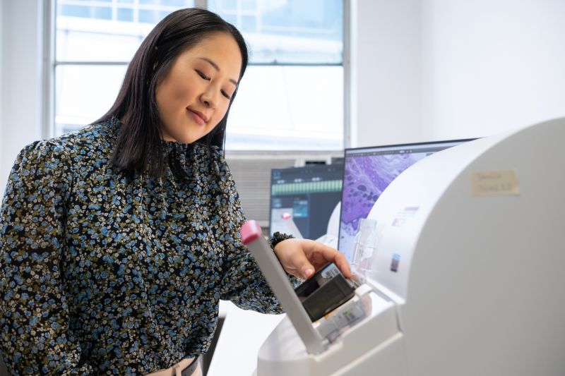
Sysmex XN1000-V Haematology Analyser
The Phenomics Australia Histopathology & Slide Scanning Service has just added the Sysmex XN1000-V haematology analyser to the service. This system delivers a highly comprehensive CBC and differential blood analysis.
As part of the comprehensive mouse necropsy evaluation, a complete blood count (CBC) and blood differential test is performed on terminal blood. This provides a general health status of the animal at end stage and other indicators that align with and support associated histopathology results.
Specifically tailored for veterinary applications, the XN1000-V is validated for mice and a number of other species, bringing a new level of precision, speed, and reliability to our service.
Investing in advanced tools like the XN1000-V is part of our ongoing commitment to excellence in research.
The analyser is available to all internal and external researchers.
Submit requests through iLab
For more information contact Tina Cardamone t.cardamone@unimelb.edu.au
Staff are always available to assist
Our Staff can provide training and assistance for independent Slide Scanner operation. Alternatively, you can simply deliver your slides to us and we will do the rest.
Standard Operating Procedures
We have a standard first line phenotyping protocol to evaluate genetically modified mouse/rat strains. Our procedures are designed to provide a thorough, first line approach assessing the entire animal.
The necropsy standard operating procedures, and the histopathology/scoring protocols were established with the assistance of consultant Pathologists and are based on International Mouse Phenotyping Resource of Standardized Screens
For standard operating procedures please contact Tina Cardamone
Location
Department Anatomy and Physiology
Level 2, Medical Building
The University of Melbourne, Parkville
Contacts for access
- Tina Cardamone: +61 3 834 48044
- Emily Gracie: +61 3 90355387
Aira Nuguid: +61 3 83448044
histopathology-scanning@lists.unimelb.edu.au
Facility hours
Monday – Friday: 8.30am to 5.00pm
Instruments are not available on weekends
Services
Clients have the option of Staff Service or unassisted instrument use after undertaking a training session. This can be arranged with Tina Cardamone or Emily Gracie.
Email: histopathology-scanning@lists.unimelb.edu.au
Training
All unassisted instrument access requires users to undertake a training session.
This is organised through the Phenomics Australia manager and incurs a fee.
Booking
Instrument reservations can be made through iLab.
Charges
Please contact histopathology-scanning@lists.unimelb.edu.au for charge schedules and quotes.
Note: Prices subject to change
Sessions may be cancelled at any time to schedule maintenance or repairs.
Notice will be issued where possible, but occasionally there will be no notice period.
The Histopathology and Digital Slide Scanning Service is supported by Phenomics Australia
The Histopathology and Digital Slide Scanning Service provides a secure interface where users are able to login and interrogate their case files and images
To access your slides on the SlideCenter viewer you will need your login details: user name and password
Access slides-SlideCenter viewer
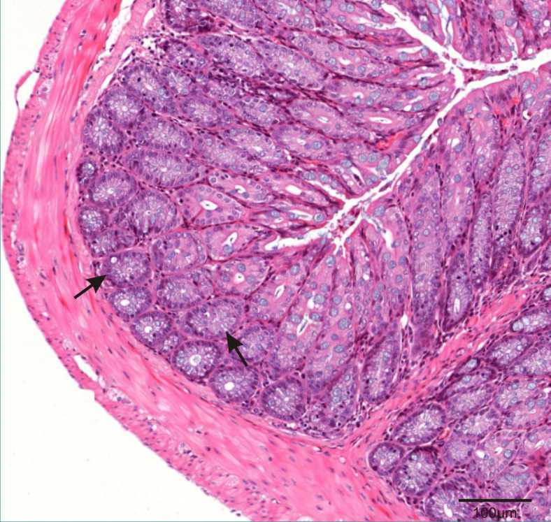
Histology sections from normal mouse tissue and organs
Clicking on an image will bring up a larger image in another tab.
-
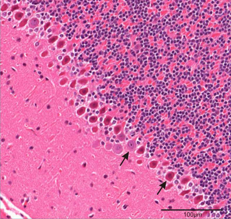
Brain - Cerebellum
Arrows: Purkinje cells -

Spinal Cord
Asterisk: Central canal
Arrows: Neurons -
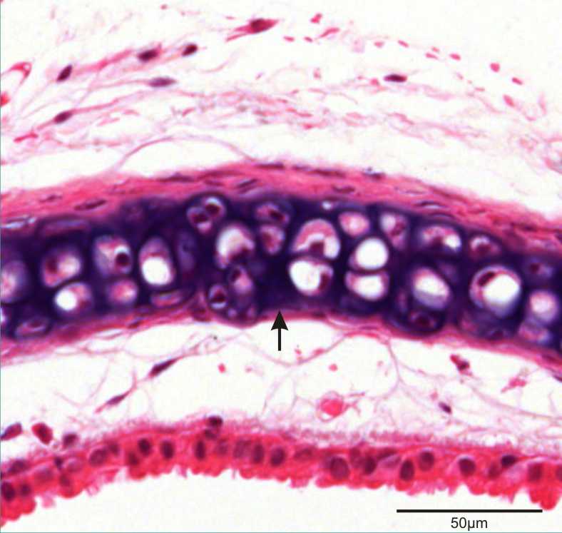
Trachea:
Arrow: Hyaline cartilage -
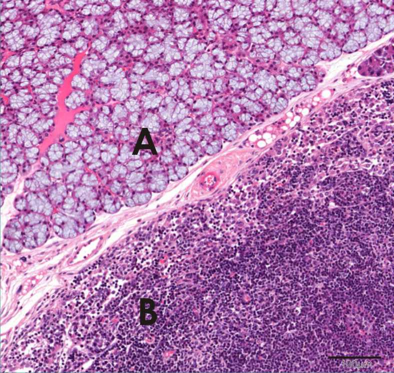
Salivary gland (A) and lymph node (B) -
Thymus
Medulla (A)
Cortex (B) -
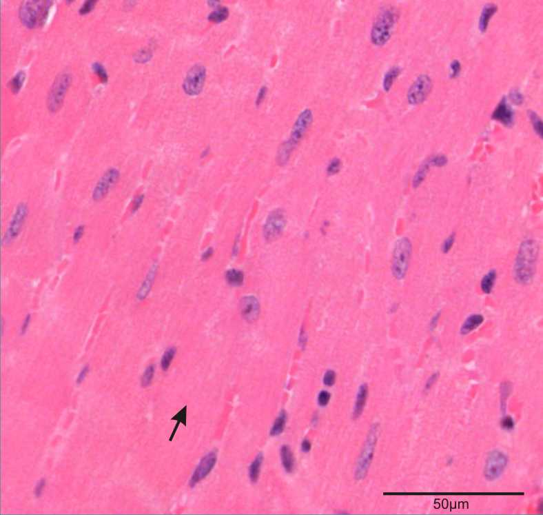
Heart
Arrow: Ventricular muscle -
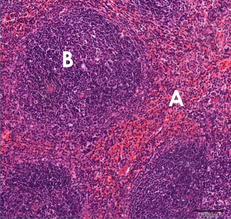
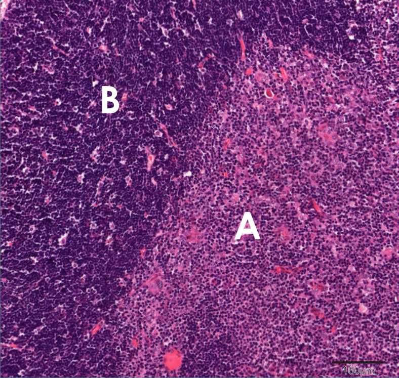
Spleen
A: Red pulp
B: White pulp -
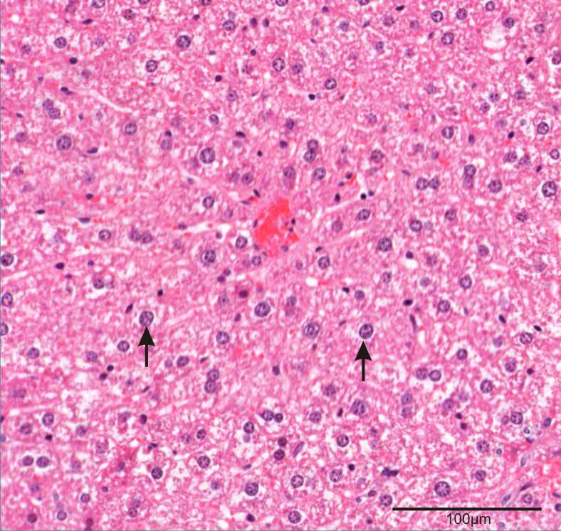
Liver
Arrows: Hepatocytes -
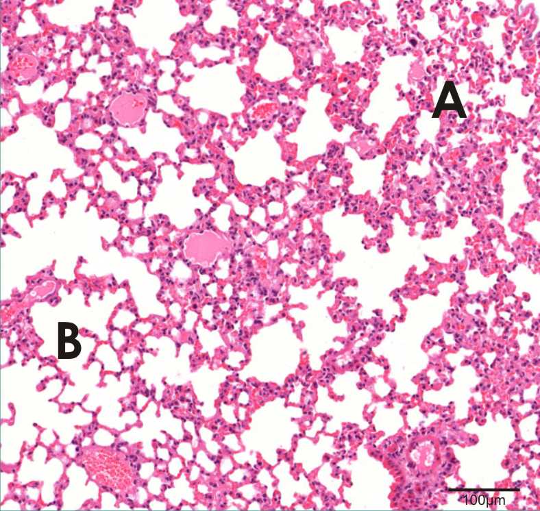
Lung
A: Alveoli
B: Alveolar ducts -
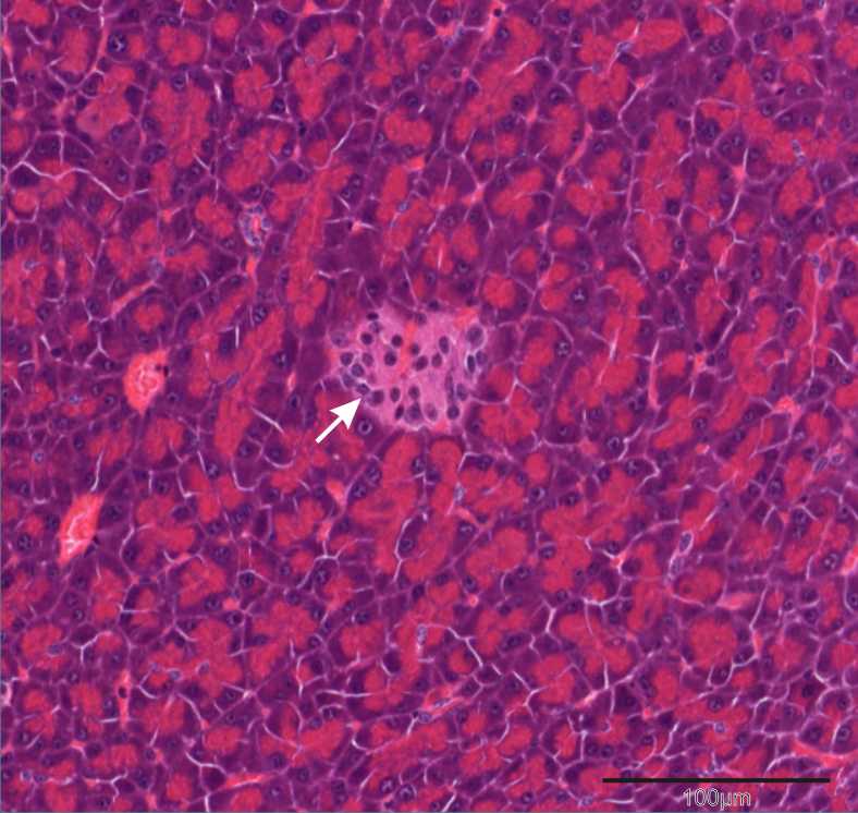
Pancreas
Arrow: Islet of Langerham -
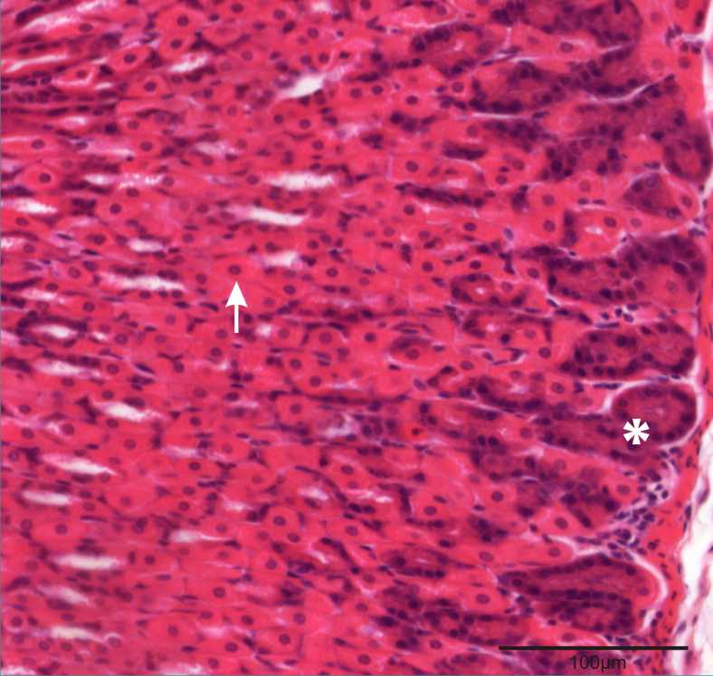
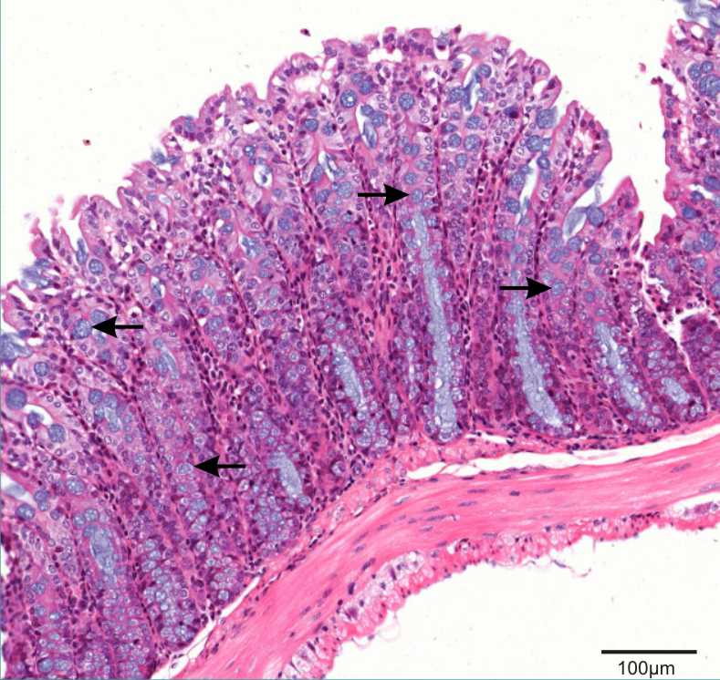
Stomach (Glandular)
Asterisk: Chief cell
Arrow: Parietal cell -
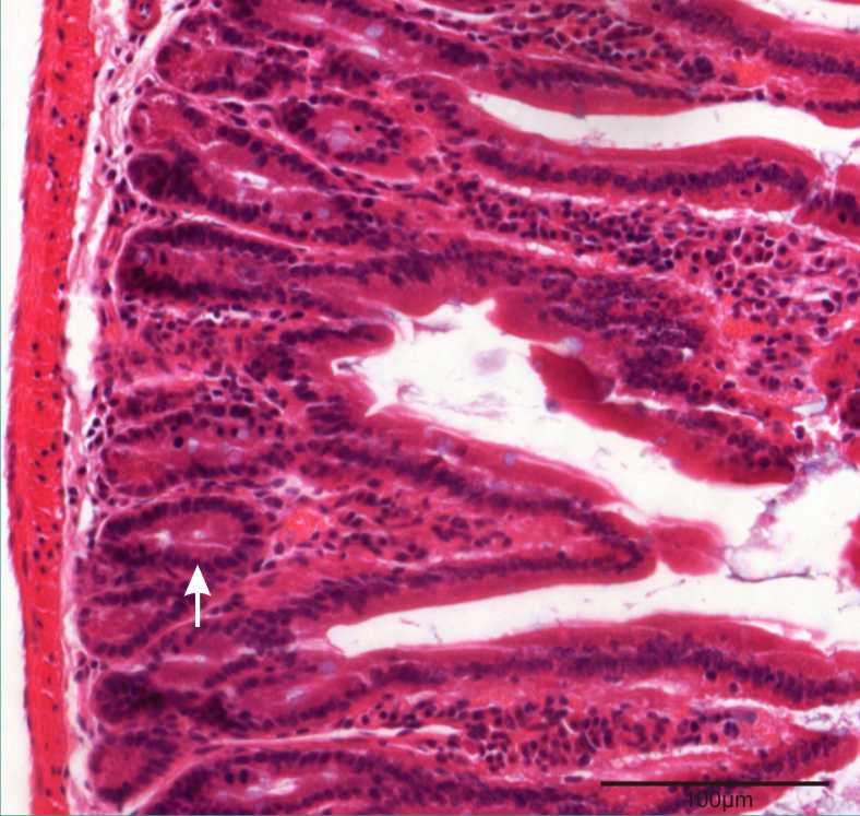
Duodenum
Arrow: Crypt of Lieberkuhn -
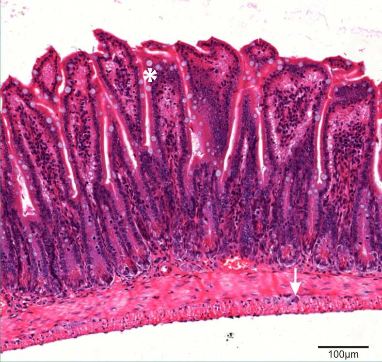
Ileum
Asterisk: Goblet cells
Arrow: Enteric neuron -
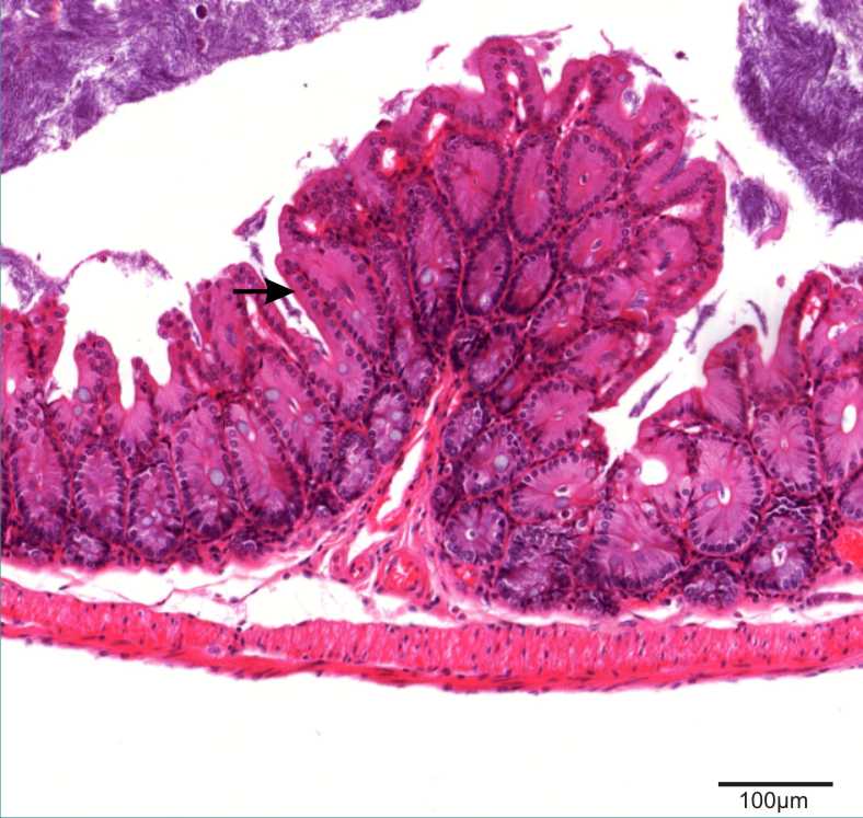
Cecum
Arrow: Columnar epithelium -
Colon
Arrows: Goblet cells -
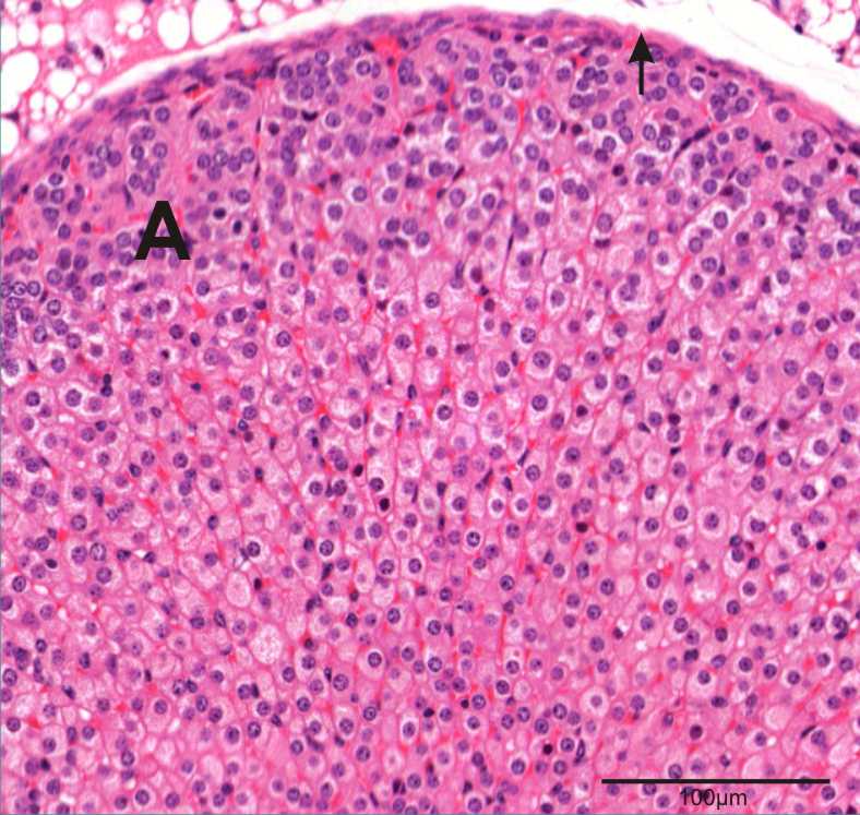
Adrenal
Arrow: Fibrous connective tissue capsule
A: Cortex region -
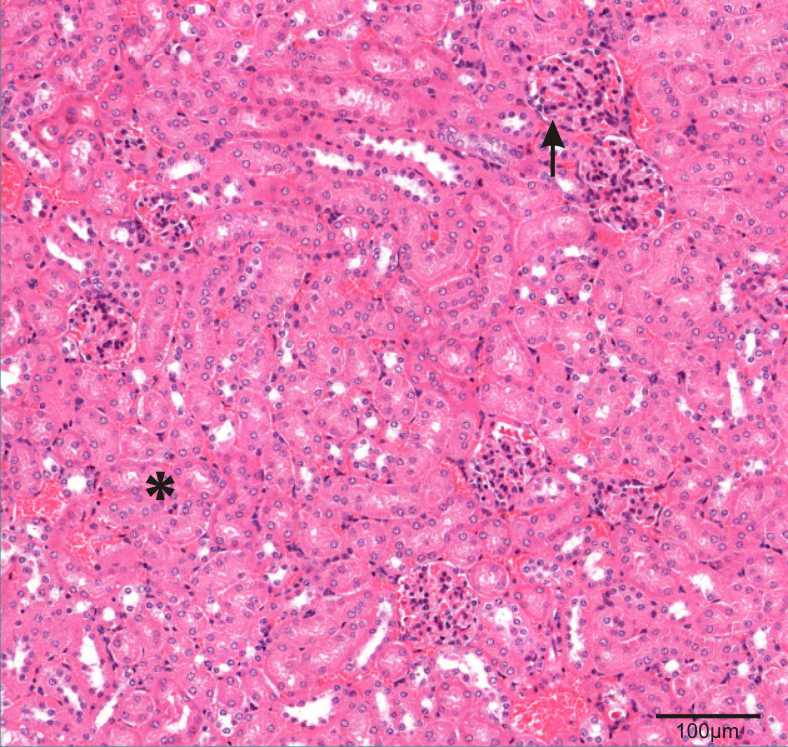
Kidney
Arrow: Glomerulus
Asterisk: Proximal convoluted tubule -
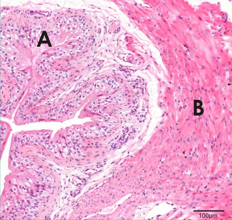
Urinary bladder
A: Transitional epithelium
B: Detrusor muscle -
Urethra
Arrows: Urethral glands -
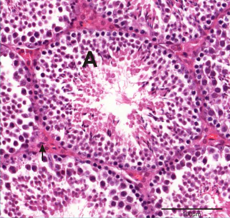
Testis
A: Seminiferous tubule
Arrow: Leydig cell -
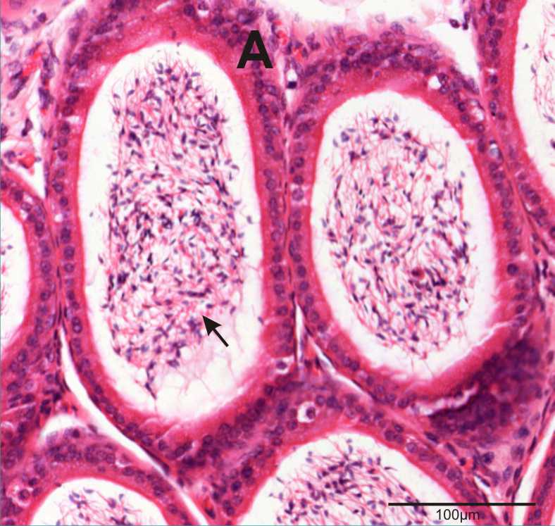
Epididymus A: Epididymal duct Arrow: Sperm -

Prostate
Arrow: Glandular component -
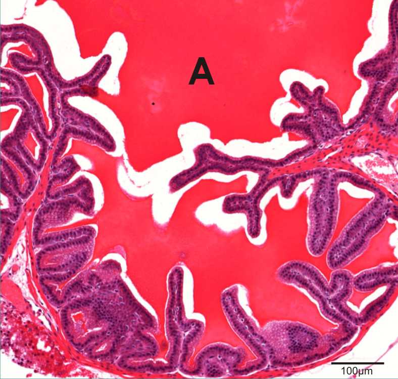
Seminal vesicles
A: Seminal fluid -
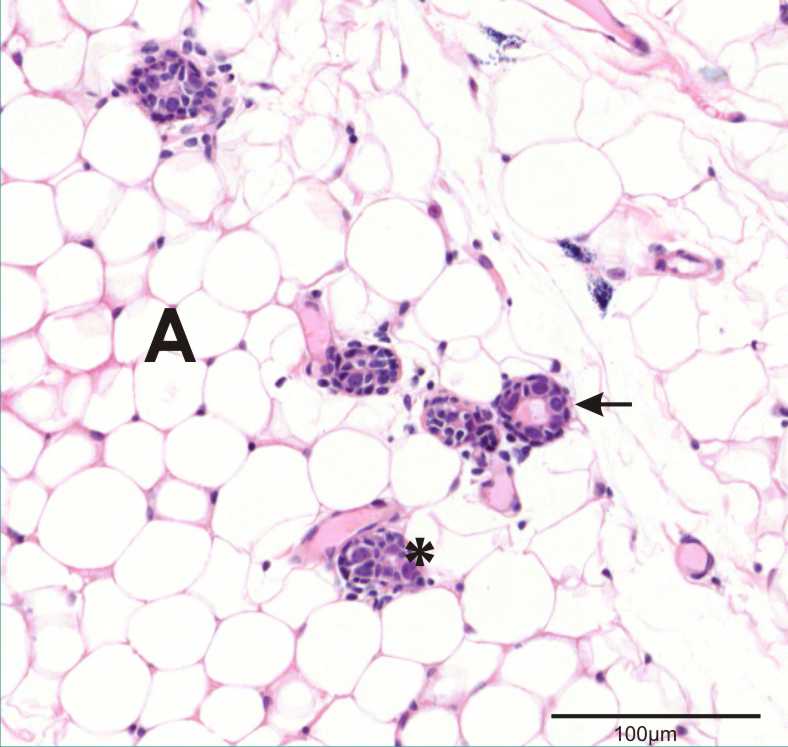
Mammary gland
Arrow: Lactiferous duct
Asterisk: Acinus
A: Adipose tissue -
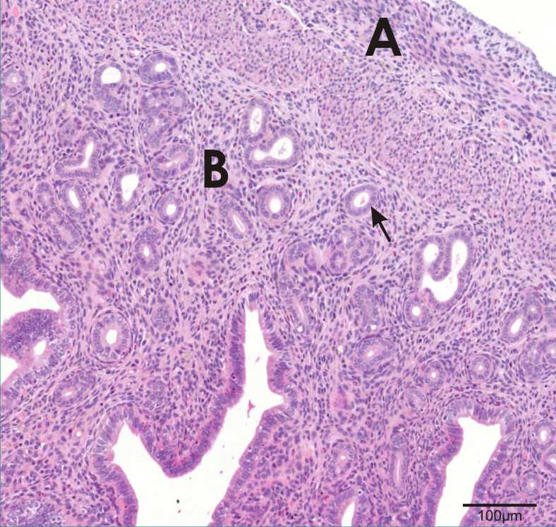
Uterus
A: Myometrium
B: Endometrium
Arrow: Endometrial gland -
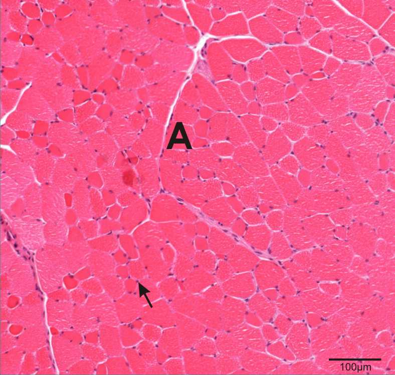
Skeletal muscle
A: Muscle fibre
Arrow: Peripherally located nuclei -

Skin
A: Epidermis
B: Dermis
C: Adipose tissue
Arrow: Hair follicle -
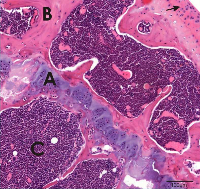
Joint of hind leg
A: Epiphyseal growth plate
B: Bone
C: Bone marrow
Arrow: hyaline cartilage -
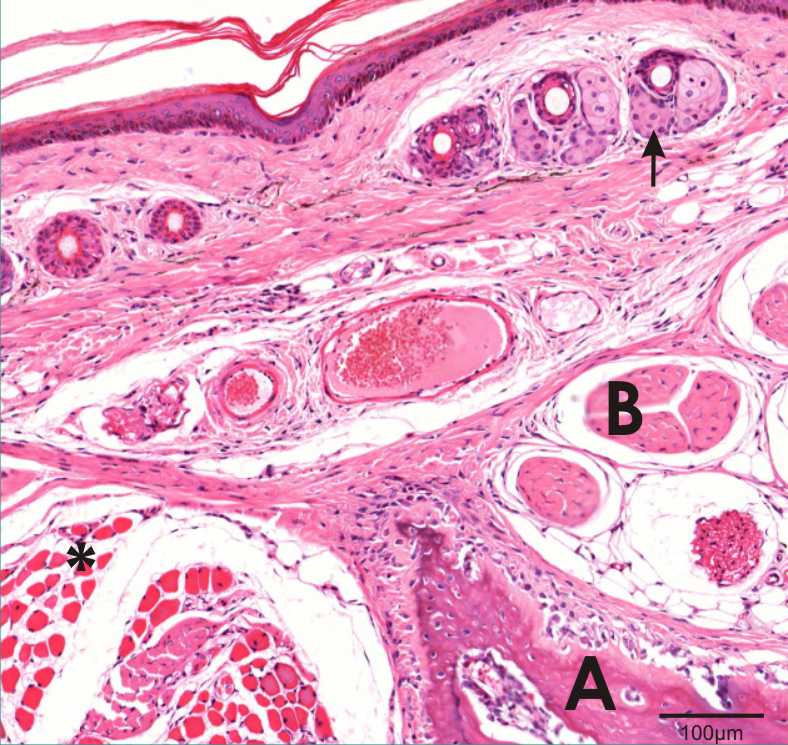
Tail
A: Vertebrum
B: Tendon
Arrow: Sebaceous gland
Asterisk: Muscle -
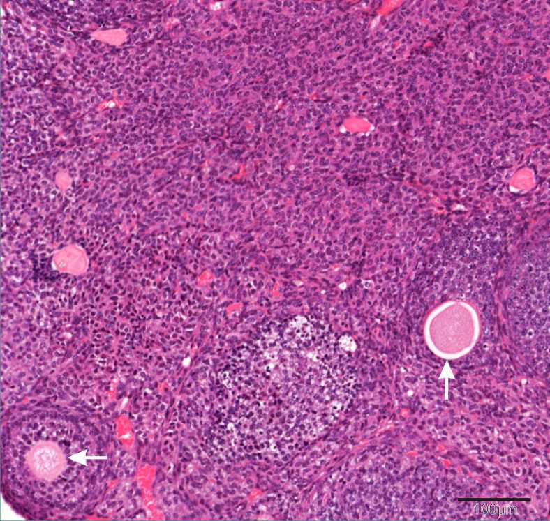
Ovary Arrows: Follicles -