'Cellular barcoding' reveals how breast cancer spreads
A cutting-edge technique called cellular barcoding has been used to tag, track and pinpoint cells responsible for the spread of breast cancer from the main tumour into the blood and other organs.
The technique also revealed how chemotherapy temporarily shrinks the number of harmful cells, rather than eliminating them, explaining how the cancer could eventually relapse.
Insights from the study, published in Nature Communications, could lead to new targeted treatments for breast cancer, the most common cancer to affect women.
Dr Delphine Merino, Dr Tom Weber, Professor Jane Visvader, Professor Geoffrey Lindeman and Dr Shalin Naik led the highly collaborative research that involved breast cancer biologists, clinician scientists, biotechnologists and computational experts at the Walter and Eliza Hall Institute of Medical Research.
Most deaths from breast cancer are caused by the metastasis, or spread, of cancerous cells from the main tumour site into other organs.
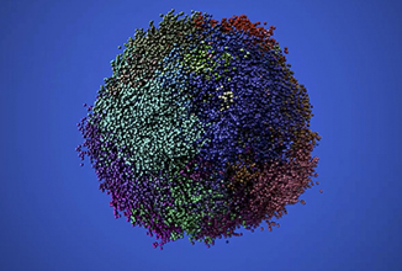
Dr Merino said the ability to pinpoint the ‘clones’ – subpopulations of cells arising from an original patient tumour – responsible for the spread of cancer was crucial for improving treatments.
“Our study revealed that only a select few clones were actually responsible for the metastasis.
“The barcoding technique enabled us to identify the clones that were able to get into the blood stream and make their way into other organs where they would ‘seed’ new tumour growth,” Dr Merino said.
Professor Visvader said the technique also allowed the researchers to see what was happening to the clones after chemotherapy was introduced.
The cellular barcoding technique was developed in 2013 by Dr Naik and Professor Ton Schumacher from the Netherlands Cancer Institute. Dr Tom Weber, a postdoctoral Fellow in the Naik lab, was in charge of the bioinformatics and barcode analysis, as well as all the figures in the paper.
Dr Naik said this new technique meant researchers could go from studying thousands of clones, to homing in on the select few variants responsible for the spread of cancer.
“Now that we know which clones are involved in the spread of breast cancer, we have the power to really focus our research to block their activity. For instance, we are curious to understand what is unique about these particular clones that enables them to successfully spread, seed and grow the cancer,” Dr Naik said.
Professor Visvader said the precision of the approach could pave the way for unravelling important mysteries in the field of breast cancer research and equip scientists with the information needed to design highly targeted treatment strategies for the prevalent disease.
“An important goal is to understand the molecular and cellular basis of how breast cancer spreads and, working with clinician scientists like Professor Lindeman, translate this knowledge from the laboratory into the clinic,” she said.
The study was supported by Australia’s National Health and Medical Research Council, the National Breast Cancer Foundation, the Joan Marshall Breast Cancer Research Fund, the Viertel Senior Medical Researcher Fellowship and the Victorian Government. Tissue for this study was provided by the Royal Melbourne Hospital Tissue Bank and the Victorian Cancer Biobank.
Credit: Walter and Eliza Hall Institute of Medical Research
Dr Shalin Naik and Dr Tom Weber are members of the Centre for Stem Cell Systems.
For more information:
Read the article.