Sea snakes
Sea snake
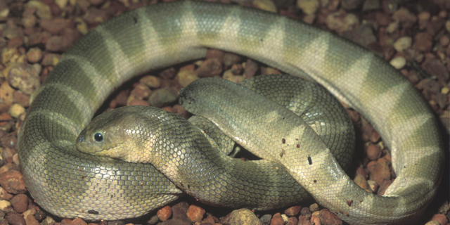
Lapemis curtus (Photo Dr Mark O'Shea)
About
Sea snakes (family Hydrophiidae) are readily identified by their flattened tails and valvular nostrils. They are, of course, excellent swimmers and divers, feeding on fish and eels. They shed their skins much more frequently than land snakes, as often as every two weeks.
The young are born alive at sea, except for those of the banded sea krait (Laticauda colubrina), which comes ashore to lay its eggs.
All sea snakes are venomous, and rhabdomyolysis is a major feature of sea snake envenomation, resulting in muscle pain, tenderness and sometimes spasm. Myoglobinuria develops after 3-6 hours. The bite itself is not particularly painful, and may go unnoticed, distinguishing it from envenomation by stinging fishes or jellyfish, both of which usually cause immediate and often excruciating pain.
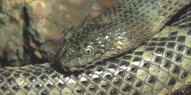
Acalyptophis peronii (Photo Dr Mark O’Shea)
Distribution
At least 32 species of sea snake have been recorded in northern Australian waters and some species are also found in the southern waters off Victoria, Tasmania and South Australia.
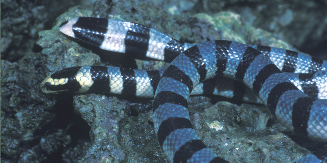
Sea krait (Laticauda colubrina; Photo Dr Mark O'Shea)
Venom
Envenomation may be treated with sea snake antivenom (based on the venom of the beaked sea snake, Enhydrina schistosa) or tiger snake antivenom. In the case of the latter, 2 ampoules should be given initially.
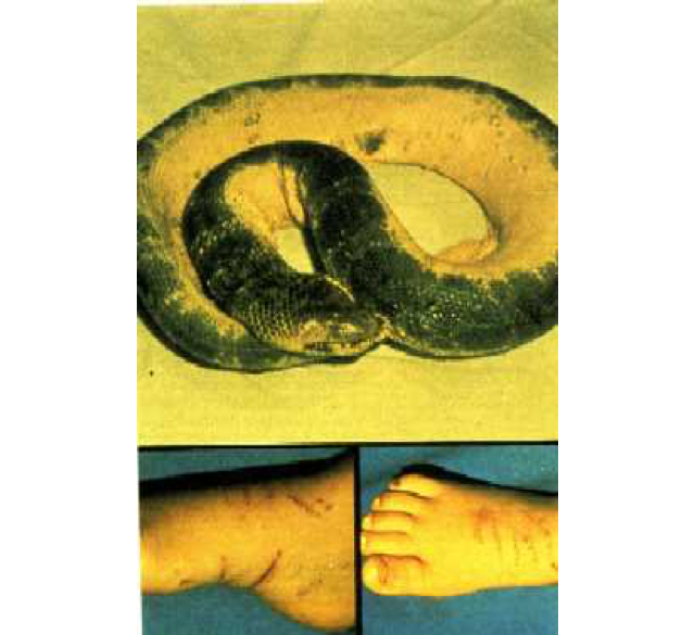
Case History: Astrotia stokesii envenomation
Believed to be the first reported case of envenomation by Astrotia stokesii in Australia.
In October 1979, a two year old girl began screaming while playing in the water at a beach 3km from Yeppoon, Queensland. A snake was noted to be wrapped around her ankle. The snake was subsequently killed and taken to Yeppoon hospital. Her mother occluded the circulation to the victim's lower limb with her hands until reaching an ambulance station. On release of the pressure, the child, who had appeared well, became drowsy and developed ptosis.
En route to hospital, she vomited and respiratory distress was evident. Twenty minutes after the bite, she was unconscious, cyanosed and in respiratory failure, and was intubated on arrival at hospital. Blood taken at this time revealed increased creatine kinase secondary to skeletal muscle damage, slightly prolonged coagulation times and a raised white cell count. The child was given three ampoules of antivenom (each 1,000 units) at 90 minutes, 2.5 hours and 3.5 hours after the bites.
Her clinical condition began to improve after the third ampoule, such that she was able to open her eyes and look around. Fourteen hours after the bite, however, her conscious state deteriorated an generalized tonic movements were observed. Three further ampoules of antivenom were administered over the next three hours, with improvement of the child's neurologic status. She was extubated at 22 hours after the bite, although she continued to experience neurologic difficulties such as incoordination, unstable gait and hallucinations until the fifth day post envenomation, at which time only the gait disturbance remained. This persisted to some degree for at least one month after the bite.
The photograph illustrates the snake involved and the appearance of the child's foot, showing multiple bite marks suggestive of severe envenomation.