The Tale of Toxicofera, part 5 (snake venom glands)
After last week’s courageous exercise in fence-sitting regarding the venomousness of varanid lizards, this week we’re jumping back onboard the anatomy train to take the gland track to…. more controversy and ambiguity. Snakes are the archetypal venomous reptiles, so you would be excused for thinking that it must be easy to agree upon which species are venomous and which aren’t, and what exactly qualifies as a snake “venom gland”. Again, thanks to the treasure trove of transitional forms that is the Toxicofera, things are not quite so simple.
Dental gland redux
If you’ve been following the Tale so far, you know that the “toxin-secreting” oral glands that the clade Toxicofera derives its name from exist in myriad forms within the clade. What seems fairly clear is that the most recent common ancestor (MRCA) of all toxicoferan lizards (including snakes) possessed “dental glands”, which are one of the synapomorphies (shared derived traits) of the clade. Since the time of that ancestor, these dental glands have diversified differently in the various toxicoferan lineages. In part 3, we had a look at the dental glands of iguanian and anguimorph lizards. We saw that the dental glands of iguanian lizards seem relatively unchanged since the time of the MRCA, but that those of anguimorph lizards are more diverse and have evolved into fully fledged “venom glands” in at least one (Helodermatidae) and possibly two (Varanidae/Lanthanotidae) lineages. In snakes, the evolution of oral glands entered warp speed in parallel with the diversification of their strategies for subduing prey without the use of limbs (imagine catching, subduing and swallowing an animal with your hands tied behind your back and your legs tied together).
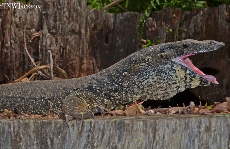
Show us your dental glands! A lace monitor (Varanus varius) says "aaaaaaaaah". This gaping beaviour is a threat display, indicating that the lizard will not hesitate to bite if it feels it cannot escape. As discussed in the previous episode of the Tale of Toxicofera, whether nor not monitor lizards should be considered "venomous", it's their teeth that do most of the damage, and this is particularly true for lace monitors, which have large, serrated teeth designed for shearing flesh. Photo: Timothy Jackson.
Evolutionary series
The diversity of oral gland anatomy amongst snakes makes deconvoluting the evolutionary history of these structures challenging but rewarding for evolutionary biologists. I’ve said this previously, but it is the preservation of “evolutionary series” of dental glands within extant toxicoferan reptiles that makes the clade such a valuable resource for researchers interested in the emergence of functional traits. Such evolutionary series should not be taken to culminate in the evolution of specialised venom systems any more than the evolutionary history of life on earth should be seen to culminate in humanity. However, when we frame the evolution of anatomical structures in relation to the existence of a particularly conspicuous functional trait – such as venom – we have the opportunity to learn a great deal about how such traits emerge. Thus, within toxicoferan reptiles we may consider the existence of a spectrum of “venom systems”, from marginal to paradigmatic cases. Paradigmatic venom systems are those which are unequivocally specialised for the production and delivery of venom, such as the “high-pressure” venom delivery systems of snakes (which we will discuss in below) or scorpions and many other venomous invertebrates. Marginal venom systems, on the other hand, may be either “incipient” or “vestigial”. An incipient venom system does not possess the function of venom production and delivery and never has but has characteristics that have been developed for that function in other lineages. The dental glands of iguanian lizards are incipient venom systems in this sense, because dental glands are the evolutionary precursors to every reptilian venom system we know about. A vestigial venom system, on the other hand, is the remnants of a formerly functional venom system that has ceased to play a role in the feeding ecology of the organism and has thus begun to decay, like the discarded old rust bucket falling into disrepair on your neighbour’s front lawn. As we shall see, vestigial venom systems are not uncommon amongst snakes.
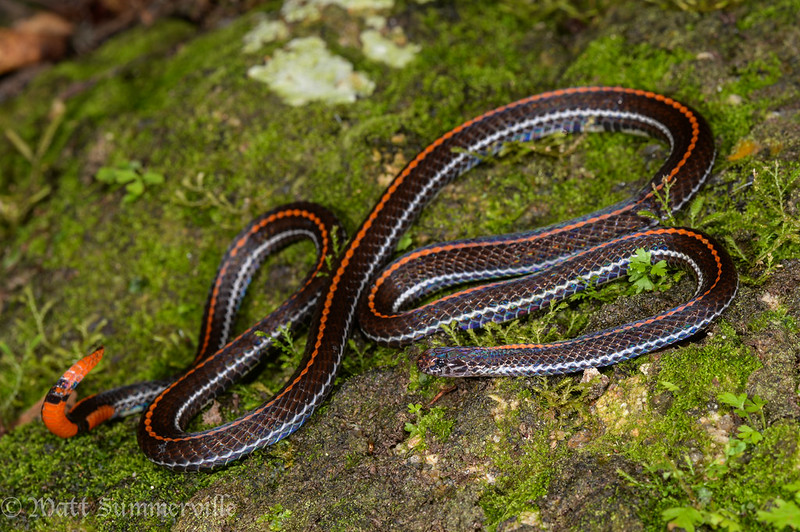
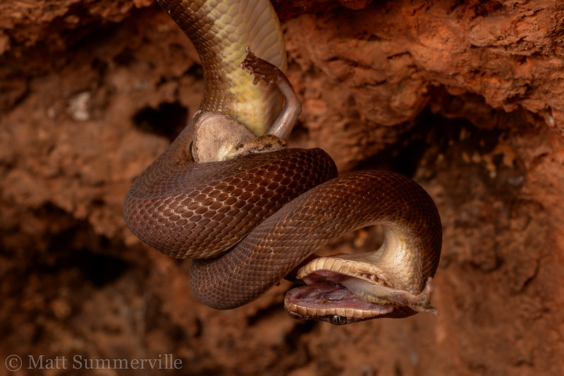 The rictal glands (see text for discussion) of pythons may be the vestiges of larger toxin-secreting oral glands possessed by ancestral snakes. Pythons, such as this Children's python (Antaresia childreni) subduing a frog, are specialised constrictors, and therefore have little need of venom to subdue their prey. Photo: Matt Summerville.
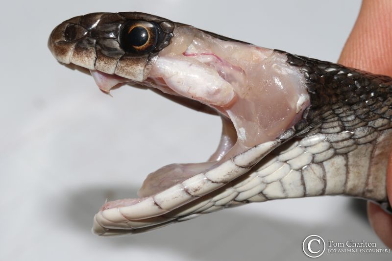
Slime factories
In contrast to anguimorph lizards (see part 3), amongst snakes it’s almost exclusively the dental gland of the top jaw (the maxillary dental gland) that has become specialised for toxin production. Typically, snake dental or labial (associated with the lips) glands of the top jaw have segregated mucous and serous (protein) secreting regions, with the mucous-secreting regions proximal (towards the nose) to the protein-secreting regions (which are under and behind the eye). In non-venomous lineages that have specialised for constriction (e.g. pythons), the protein-secreting portion of the glands has been reduced almost to nothing, and the large “labial glands” secrete mucous almost exclusively. This capacity for producing a surfeit of slime is important for these snakes that are able to subdue exceptionally large prey and are unable (like almost all snakes) to break the prey apart. Swallowing a large furry mammal whole requires a lot of lubrication, which the labial glands supply. The protein-secreting region of the gland that remains is located right at the back of the upper jaw, where it hinges with the lower jaw. These glands are known as “rictal glands” and are not exclusively possessed by constricting snakes. In fact, they are widely distributed and vary a great deal in size – often they are very small, but not always.
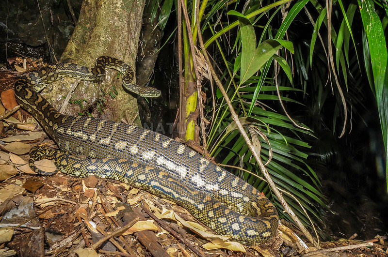
Pythons, like this carpet python (Morelia spilota) can swallow huge meals. Labial glands that secrete large quantities of mucous are an essential part of this process, as the mucous lubricates the meal, making it easier to swallow. Photo: Scott Eipper.
Ancestral snake strategies
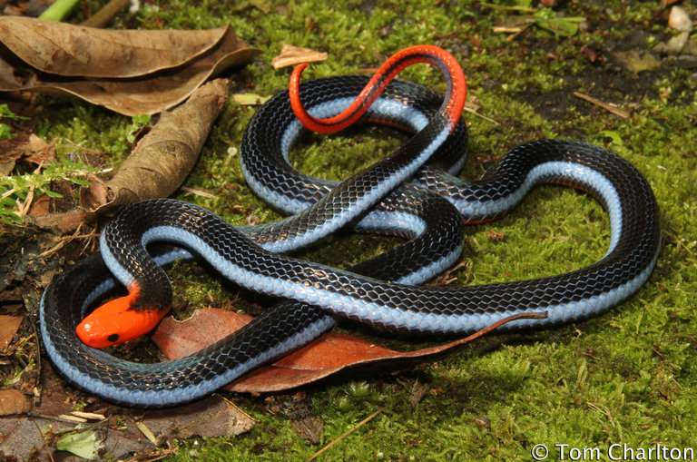
Some species have both a “supralabial venom gland” and a rictal gland, others have one but not the other. In a few of the most “plesiomorphic” (similar to an inferred ancestral state) snake lineages, the rictal gland is very large and the venom gland is reduced or absent. In at least one such snake – the red-tailed pipe snake (Cylindrophis ruffus) of Southeast Asia, the rictal gland expresses genes that encode neurotoxins in other snake lineages. Intriguingly, the tiny rictal glands of non-venomous constrictors also express genes encoding neurotoxins, and the secretions of these glands are toxic when injected into laboratory rodents. This doesn’t necessarily mean that these snakes are “venomous”, as the secretions of laboratory rodent salivary glands are also toxic when injected into laboratory rodents. However, it does provide yet more interesting evidence that can help us deconvolute the evolutionary history of snake venom systems (if it doesn’t simply confuse us further). What seems clear is that the rictal glands were probably once part of a single maxillary dental gland in some ancestral snake that is lost to the sands of time. This ancestral snake may have been at least marginally venomous, deploying its toxin-rich oral secretions in combination with grappling/constriction/biting hard as part of a combined prey subjugation strategy, similar to that which is utilised by many extant snakes. Why some lineages have favoured the rictal gland over the venom gland and vice versa, and why others have retained both structures, is a question we still don’t have an answer to.
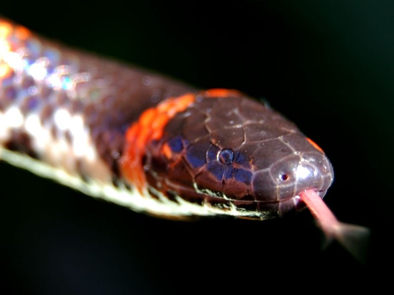
The red-tailed pipe snake (Cylindrophis ruffus) is believed by some researchers to be similar in form to the ancestor of all snakes. This species has a large toxin-secreting rictal gland and subdues its prey by biting it repeatedly and grappling with in (but not constricting). Is it "venomous"? We don't know. Photo: Wikimedia Commons.
Venom glands
We could quite easily expend several thousand more words discussing variations on these themes, but let’s skip that and just go straight to the good stuff – the venom gland itself. Broadly speaking, the venom gland is the synapomorphy of the so-called “advanced snakes” (Caenophidia, Colubroidea, or Colubroides, depending on your preferred nomenclature). By now, it will come as no surprise to hear that venom glands come in all shapes and sizes within this spectacularly diverse clade, which contains three quarters of the world’s species of snake. It is probably also predictable that this diversity in form has generated controversy surrounding which glands should be granted the title “venom gland”. My personal preference is to keep things as simple as possible (since snake oral gland nomenclature is tediously complex at the best of times) and call the post-orbital (behind/beneath the eye) supralabial (associated with the upper lip) maxillary (of the upper jaw) protein-secreting dental gland of advanced snakes a “venom gland” wherever it occurs (so you can ignore all those other words). This may seem to contradict the prevarication about which species are venomous that has characterised previous episodes of this Tale, but here I am choosing to avoid arbitrarily complicating things by naming the gland after its most well-characterised function, rather than trying to draw circles around those most likely to be specialised for that function and excluding those which may not be. Previously, the name “Duvernoy’s gland” was used for this gland in non-front-fanged snakes and “venom gland” was reserved for front-fanged species. Personally, I feel this scheme only confuses things further because the name “Duvernoy’s gland” is not usefully descriptive of form or function, the “venom glands” of front-fanged snakes evolved their similar features independently, and many non-front-fanged snakes are quite clearly venomous. This debate about appropriate naming of homologous structures (structures arising from a common ancestor) is ongoing and is another example of the difficulty we have in suppressing our desires to carve nature into clearly defined categories. No matter which naming scheme is adopted, this dream cannot be fulfilled – there are simply too many shades of grey – which recommends usage of the most inclusive terms that can reasonably be justified. We must move on but please see this paper for further details.
The large venom gland of this Papuan taipan (Oxyuranus scutellatus) is located "post-orbitally" (behind the eye) and is associated with the upper lip ("supralabial") and upper jaw ("maxillary"). Many terms have been used for this gland in its various forms, but I prefer to avoid the jargon and simply call it a "venom gland" , wherever it occurs. Photo: Tom Charlton.
There are three lineages of front-fanged venomous snakes, each of which evolved the front-fanged condition independently. These are the family Elapidae (cobras, taipans, and their relatives), the family Viperidae (vipers and pit vipers) and the clade containing the genera Atractaspis and Homoroselaps (stiletto snakes/mole vipers and harlequin snakes). As mentioned, the venom glands of these three lineages share certain features, which are associated with their being components of a “high-pressure” venom delivery system. The most notable of these is the attachment of compressor musculature which squeezes the gland during a bite (or a spit, in the case of spitting cobras), forcefully propelling the venom down the duct and through the hollow fang. The entire system is something like a hypodermic needle. These shared features evolved convergently, however, and the glands are different in many respects, including which muscles attach to them. For this reason, these glands are “homoplasic” (convergent in form and function), but the features which distinguish them from the venom glands of non-front-fanged snakes are not homologous (descended from a common ancestor). In fact, evolving compressor musculature is such a neat trick that it has occurred several times amongst the non-front-fanged snakes too, albeit in a far less effective form – yet again, we seem to be catching evolution in the act by uncovering incipient forms of particular structures.
Extravagant extensions
 Incredibly, the venom glands of this banded coral snake (Calliophis intestinalis) extend well beyond the skull and down into the body cavity, for as much as a quarter of the snake's body length! "Long-glanded" snakes exist in all three of the front-fanged clades, indicating that this condition has evolved multiple times independently (including two occasions within the family Elapidae).....nobody really knows why. Photo: Matt Summerville.
The venom glands of non-front-fanged snakes vary considerably in size and whether or not they possess a “lumen” (a space in the centre of the gland for the storage of secreted venom). The size of the gland, along with the size of the associated teeth and whether or not they are grooved (which, by the way, also varies a lot!) is not always correlated with the potency of the snake’s venom or its apparent importance in their feeding ecology. Indeed, the venom glands of many non-front-fanged snakes are considerably larger than those of many front-fanged elapid snakes, and those of the latter often lack a lumen despite their obvious importance in the feeding ecology of the snakes that possess them. Venom gland morphology is generally conserved within each of the front-fanged lineages, but there are some weird and wonderful examples that buck this trend. “Long-glanded” forms, in which the venom gland extends well beyond the head and into the body cavity, for as much as a quarter of the length of the snake (!) are found in each lineage. They tend to be possessed by burrowing species with relatively slender heads, but many closely related burrowing species lack them, so the evolutionary “justification” for such extravagantly large glands remains unclear. It may simply be the case that it only takes a small genetic change to cause the glandular tissue to break its usual bounds in this matter, and if there is even a slight selective advantage (and no significant penalty) to the possession of such glands then….why not? More is more!
 Like its cousin the banded coral snake (pictured above), the spectacular blue coral snake (Calliophis bivirgatus) possesses extravagantly extended venom glands.
Use it or lose it
The mosaic sea snake (Aipysurus mosaicus) feeds on fish eggs. As it apparently no longer has a need for venom, its venom glands and fangs are greatly reduced, to the point of being "vestigial". Photo: Dave Gower.
Vestigial venom glands are also common amongst non-front-fanged snakes, and there are even a few examples amongst the elapids. Numerous non-front-fanged lineages have become specialised constrictors and seem to no longer have a need for venom to subdue their prey. Accordingly, their venom glands shrink in size almost to the point of disappearance (and occasionally do seem to disappear altogether), and the mucous-secreting region often extends to fill the vacant real estate along the top jaw. Similar things happen when snakes start feeding on “defenceless” prey, such as the eggs of fishes, frogs, birds, or other reptiles. Egg-eaters occur in many non-front-fanged lineages, and there are a number of elapid snakes that have also taken this route to easy living. Interestingly, the defensive function for which many snakes utilise their venom seems insufficiently beneficial for these snakes to maintain a functional venom system, and it often withers away – “use it or lose it” is a basic evolutionary principle. Front-fanged elapid snakes with apparently vestigial venom systems include sea snakes that feed exclusively on fish eggs. This behaviour, and the associated reduction of the venom system, has evolved multiple times independently amongst marine elapids. Perhaps even more surprising is the evidence that certain lineages may have transitioned back-and-forth, with essentially non-venomous snakes evolving from venomous species and then reverting to the functionally venomous condition. In evolution, whatever can happen, will happen!
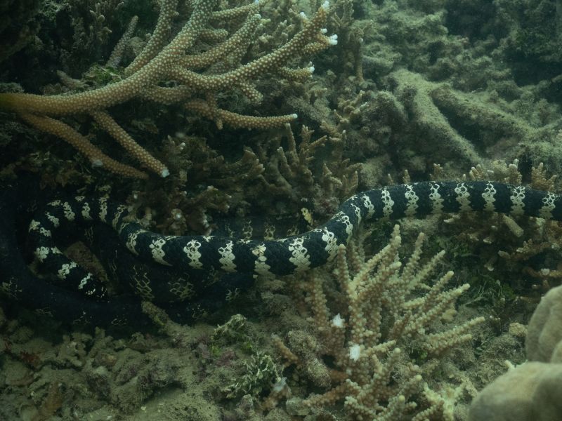
The turtle-headed sea snake (Emydocephalus annulatus) is also an egg-eater with reduced venom glands, a condition it has evolved independently from the mosaic sea snake (picture above). Photo: Jenna Crowe-Riddell.
Exemplifying the plethora of possible forms of snake venom glands are the species of snake that think they are anguimorph lizards and have evolved toxin-secreting dental glands (functional “venom glands”) on their lower jaw. These snakes, members of the genus Dipsas, are found in the tropical Americas, and possess the typical venom gland of the upper jaw as well as their unique mandibular arrangement. They insert their weaponised lower jaws into the shells of the snails they feed upon, envenoming the molluscs to make them easier to extract (and possibly to reduce their production of slime).
There are more species of reptile than any other kind of back-boned animal – reptiles are the most speciose lineage of vertebrates on the planet. Some might quibble and say there are slightly more species of birds than reptiles, but readers of this blog (and people interested in phylogenetics) know that birds are reptiles. In turn, Squamata (snakes and lizards) is by far the most speciose extant lineage of non-avian reptiles. In fact, over 96% of all reptile species alive (and described) on the planet today are members of this order. Snakes are (again, by some margin) the most speciose group of squamates, and specialised oral glands (including, but not limited to, venom glands) are a big factor in the success of these limbless wonders. When this fact is considered alongside the diversity of snake lifestyles, it becomes unsurprising that snake oral gland anatomy is a complicated subject. We’ve only scratched the surface here, but hopefully we’ve covered enough to reinforce the central point of this series on Toxicofera – the richness of transitional forms within the clade makes any attempt to draw lines in the sand unequivocally dividing the “venomous” from the “non-venomous”, the “venom glands” from the “Duvernoy’s glands” a hopeless endeavour. Again, categorising things is a useful aid to investigating them, but strictly defining these categories is a far less interesting and important task than taking advantage of the opportunity the grey areas present for us to gain insight into the many-branching path traversed by organismal lineages over evolutionary time.
Tune in next week for more evolutionary thinking!
- Timothy