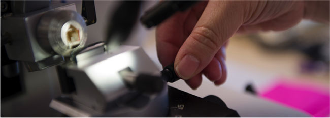
Microscopy
The Department of Microbiology and Immunology has facilities for light and epifluorescent, confocal and multi-photon microscopy, as well as tissue sectioning. The confocal microscopes are part of the Biological Optical Microscopy Platform (BOMP). Training is essential before any of these instruments can be used.
Light and Epifluorescence
The Department is equipped with three microscopes capable of epifluorescent & light microscopy.
- Leica DM-IL HC (Inverted)_Wavelength: 380 - 780 nm (VIS range, Hg lamp)
- Leica DM LB HC (upright)_Wavelength: 380 - 780 nm (VIS range, Hg lamp)
- Leica DMI 4000 B (Inverted)_Wavelength: 380 - 780 nm (VIS range, Hg lamp)
All systems run using LAS AF software (Leica) and two cameras are available for image acquisition.
- DC 400- Colour camera, 8 Megapixel- suitable for light microscopy, fluorescence microscopy and time lapse and movies.
- DC350-Black and white camera, 8 megapixel. Superior senstivity for fluorescence microscopy.
ACCESS:
Training is required before using these instruments.
FACILITY:
Room 7020
CONTACT:
Manager: Adam Lopez-Denman
Office: 7006
Lab: 7028
Email: adam.lopez@unimelb.edu.au
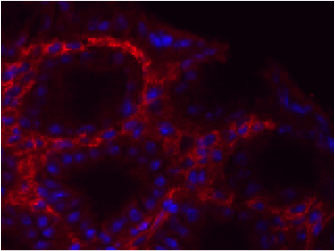
Confocal
The Department has two confocal microscopes that are maintained by the Biological Optical Microscopy Platform (BOMP). Training, assisted use and advice can be provided by contacting BOMP.
- Zeiss LSM700: Inverted stand, 4 laser lines, two fluorescence detectors, one transmitted light detector, motorised stage (z stack & tile scan), incubation chamber for live cell imaging experiments.
- Zeiss LSM710: Inverted stand, 6 laser lines, with 34 channel spectral detector, two high sensitivity (GaAsP) detectors and motorised stage functions (z stack & tile scan).
Once trained the systems are bookable from the following booking system.
Costs (correct as of July 2014)
| Un-assisted use (DMI) | Free |
| Training/assisted use (DMI) | Free |
| Un-assisted use (Other UoM) | $35/hr |
| Training/assisted use (Other UoM) | $90/hr |
| Un-assisted use (External) | $50/hr |
| Training/assisted use (External) | $90/hr |
FACILITY:
Room: 7020
BOMP GENERAL CONTACT:
Tel: 9035 3021
Email: bomp-enquiries@unimelb.edu.au
Web: www.microscopy.unimelb.edu.au
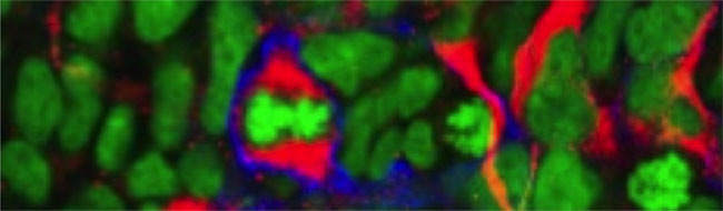
Multiphoton
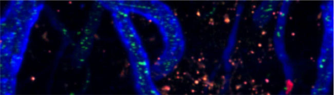
Associate Professor Scott Mueller and Professor William Heath manage the multi-photon facility for the Department of Microbiology and Immunology. The facility has two multi-photon microscopes (Zeiss 710 NLO & Olympus FVMPERS) designed to facilitate intravital imaging of mouse tissues and organs and have significantly expanded the possibilities for research in microbiology and the immune system.
Training and support are provided. For further details contact either Associate Professor Scott Mueller or Professor William Heath.
ACCESS:
Via collaboration with the Mueller & Heath labs.
CONTACTS:
Associate Professor Scott Mueller
Email: smue@unimelb.edu.au
Professor William Heath
Email: wrheath@unimelb.edu.au
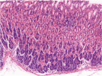
Tissue Sectioning
Frozen tissue sectioning: Leica CM3050S Cryostat
- For OCT-embedded tissue sectioning
- From 2um to 100um thickness
- Chamber temperature from -25C to -10C
- Ideal for immunofluorescence / confocal microscopy (unfixed, PFA-fixed tissue)
Fresh tissue sectioning: Leica VT1200S Vibratome
- For fresh / live tissue sectioning
- Fully automated vibrating blade
- Vibrocheck(TM) calibration to correct blade deflection
- Ideal for whole-mount microscopy (confocal / 2-photon)
- Suitable for live explant imaging (confocal / 2-photon)
ACCESS:
For access and training, contact Associate Professor Scott Mueller.